
Written by Vasileios Skouteris MD, FEBO, Ophthalmic Surgeon
Page contents
- What is the cornea and why is its inflammation serious?
- Infectious keratitis
- Non-infectious keratitis
- Symptoms
- Who is most at risk?
- Diagnosis
- Treatment
- What to do until you see an ophthalmologist
- Keratitis in children
- How long does treatment take?
- Complications
- Prevention
- When to get examined
- Frequently Asked Questions about Keratitis
Keratitis is inflammation of the cornea, the clear “window” at the front of the eye. It causes pain, marked redness, sensitivity to light and blurred vision, and in its infectious forms it can leave a permanent scar within a few days. The most common cause of serious keratitis is improper contact lens use. Any red, painful eye in someone who wears contact lenses should be examined the same day.
What is the cornea and why is its inflammation serious?
The cornea is the clear dome that covers the iris and the pupil. It is about half a millimetre thick and has two key roles: it protects the inside of the eye and provides two thirds of its focusing power, which makes it the most powerful “lens” in our visual system. To do its job it must be perfectly clear and smooth.
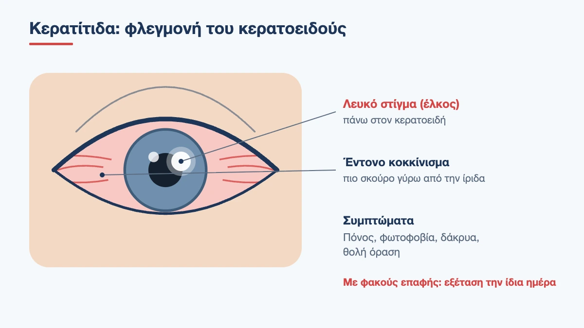
Two of its special features explain why keratitis is so painful and why it is dangerous. First, it is the most densely innervated tissue in the body, with hundreds of times more nerve endings than the skin. Even a tiny abrasion causes disproportionate pain. Second, it has no blood vessels, because they would spoil its transparency. This means that the body’s defences arrive slowly and that healing happens with scar tissue, which is opaque. A scar in the centre of the cornea works like a smudge on a camera lens.
The outer layer, the epithelium, is the barrier against microbes. As long as it is intact, infection is difficult. Anything that injures or weakens it, from a contact lens worn for too many hours to a twig in the garden, opens the door.
Infectious keratitis
Bacterial keratitis
This is the most common form of infection and the one that progresses fastest. It starts with pain and redness, and within 24 to 48 hours a white or yellowish spot appears on the cornea, the ulcer, together with a pus-like discharge. In contact lens wearers the most common culprit is Pseudomonas, a bacterium that lives in water and in lens cases and can melt the cornea within two to three days. In people who do not wear lenses there is usually a preceding injury, severe dry eye or an eyelid problem, and the usual organisms are Staphylococcus and Streptococcus.
Herpetic keratitis
This is caused by the herpes simplex virus, the same virus that causes cold sores. The virus stays dormant in the nerve and reactivates from time to time, often after a fever, intense stress, sun exposure or lowered immunity. It almost always affects one eye and produces a characteristic branching ulcer, like the branch of a tree, which shows up with a special dye. Its main problem is recurrence: each episode can leave a little more haze and reduce the sensitivity of the cornea. A related condition is keratitis from the herpes zoster virus, when shingles appears on the forehead and around the eye.
Adenoviral keratitis
This often follows viral conjunctivitis. One to two weeks after the redness, small round opacities appear under the epithelium, causing hazy vision and sensitivity to light. They are not dangerous, but they can persist for months.
Fungal keratitis
This is less common but difficult to treat. The typical history is an injury with plant material, such as a twig, leaf or thorn, during farm work or gardening. It progresses more slowly than bacterial keratitis, often with less pain at first, which is why diagnosis is delayed. Treatment lasts from weeks to months.
Acanthamoeba keratitis
Acanthamoeba is a protozoan that lives in tap water, swimming pools, the sea, hot tubs and soil. Almost all cases involve contact lens wearers who swam or showered in their lenses, or rinsed their lenses or case with tap water. Its hallmark is very severe pain, out of proportion to how the eye looks in the early stages. It is one of the most difficult eye infections, with treatment lasting many months. You can find out more in the article on contact lenses at the beach and in the pool.
Book an appointment with the doctor
An examination and clear advice on your case at our practice in Neo Psychiko.
Non-infectious keratitis
Not all keratitis is caused by a microbe. Equally common are the forms in which the cornea becomes inflamed because it has been injured, has dried out or has been attacked by the immune system.
- Photokeratitis: a burn of the epithelium from ultraviolet radiation. It appears 6 to 12 hours after welding without a mask, after many hours in snow or at the sea without sunglasses, or from sunbeds. It is very painful, but heals completely in 1 to 2 days
- Exposure keratitis: when the eyelids do not close properly, for example in facial nerve palsy, proptosis or after surgery, the lower part of the cornea dries out and becomes damaged
- Dry eye keratitis: in severe dry eye the epithelium becomes covered in tiny punctate lesions, causing burning and blurring that changes with blinking
- Marginal keratitis from blepharitis: a hypersensitivity reaction to the toxins of the Staphylococcus that lives on the eyelids. It produces small infiltrates at the edge of the cornea and tends to recur
- Neurotrophic keratitis: when the cornea has lost its sensation, after herpes, shingles, diabetes or neurosurgery, it does not heal properly. The paradox is that it does not hurt, which is what makes it insidious
- Autoimmune keratitis: in rheumatoid arthritis and other systemic diseases, the cornea can thin at the periphery. It requires close collaboration with a rheumatologist
- Toxic keratitis: from excessive or long-term use of eye drops, especially those containing preservatives, or from misuse of anaesthetic drops
- Contact lens overwear keratitis: the lens deprives the cornea of oxygen. Sleeping in lenses or wearing them for too many hours causes a red, painful eye in the morning, even without infection
Symptoms
- pain, from a gritty feeling to sharp, piercing pain that stops you opening the eye
- marked redness, darker around the iris
- photophobia, that is, pain in bright light
- watering and spasm of the eyelids
- blurred vision
- discharge, watery or pus-like
- a white or grey spot on the cornea, which can sometimes be seen in the mirror
- a feeling that there is a foreign body that does not go away with rinsing
How does it differ from conjunctivitis?
In simple conjunctivitis the eye is red, stings and has sticky discharge, but vision is good, light is not particularly bothersome and the pain is mild. When there is real pain, photophobia or reduced vision, the damage is deeper and the cornea must be checked. So three things raise the alarm level in a red eye: pain, light sensitivity and blurred vision.
Who is most at risk?
The most important risk factor for infectious keratitis is contact lens wear. The risk multiplies with sleeping in lenses, contact with water, wearing them beyond their replacement date and poor care of the lens case. Other risk factors include:
- injury to the cornea, even a minor one
- severe dry eye and eyelid disorders
- a previous episode of herpes in the eye
- long-term use of steroid eye drops
- diabetes mellitus and immunosuppression
- previous corneal surgery
- outdoor work or work with plants without protective glasses
Diagnosis
Diagnosis is made at the slit lamp, the ophthalmologist’s microscope. A drop of yellow dye, fluorescein, stains the areas where the epithelium is missing and, under blue light, reveals the shape and size of the lesion. The shape often gives away the cause: dendritic (branching) in herpes, round with a white infiltrate in bacterial infection, ring-shaped in Acanthamoeba, and scattered dots in dry eye and UV burns.
We also check for inflammation inside the eye, measure the eye pressure and test corneal sensation, which is reduced in herpes. We turn the upper eyelid over to rule out a foreign body hidden underneath.
When the ulcer is large, central or not responding to treatment, a sample is taken from its surface for culture, to identify the organism and test which medicines it is sensitive to. The sample is taken after an anaesthetic drop and is painless. If you wear contact lenses, bring your lenses, case and solution with you: they can be cultured too.
Treatment
Treatment depends entirely on the cause, and this is why self-treatment is dangerous. A medicine that helps in one form can destroy the eye in another.
Bacterial
Antibiotic drops on a very intensive schedule, hourly at first, and even through the night during the first 24 hours for severe ulcers. As the infection settles, the frequency is gradually reduced. Drops that dilate the pupil are added to reduce the pain caused by spasm of the internal eye muscles. The eye is not covered with a pad, because warmth and moisture favour bacteria. The patient is re-examined daily for the first few days.
Herpetic
Antiviral medication, as a gel in the eye or as tablets by mouth, for 2 to 3 weeks. When the inflammation affects the deeper layers, steroid drops are added, always under antiviral cover and with close monitoring. People with frequent recurrences are given preventive tablets for months, which reduces the risk of a new episode by about half. More details in the article on herpes in the eye.
Fungal and Acanthamoeba
Special antifungal or anti-amoebic drops, often specially compounded, on an intensive schedule for weeks and then less frequently for months. These organisms form resistant forms within the cornea, and stopping treatment early leads to recurrence. Patience and strict adherence to the schedule are essential.
Non-infectious forms
- Photokeratitis: lubricants, antibiotic ointment for protection, painkillers by mouth and rest in a dark room. It settles in 24 to 48 hours
- Dry eye and exposure: frequent preservative-free artificial tears, gel or ointment at night, taping the eye closed during sleep and treating the underlying cause
- Marginal keratitis from blepharitis: a short course of steroid with antibiotic and, above all, daily eyelid hygiene to stop it coming back
- Neurotrophic: intensive lubrication, a bandage contact lens, drops made from the patient’s own serum and, for persistent ulcers, an amniotic membrane graft
- Contact lens related: stopping lens wear until fully healed and reviewing the lens type and how they are used
Three things you should not do
- Do not use steroid drops you find at home. In a herpetic, fungal or amoebic ulcer, steroids without proper cover dramatically accelerate the damage
- Do not use anaesthetic drops for relief. They prevent healing and hide any worsening
- Do not put your contact lenses back in on an irritated eye “to see if it has passed”
What to do until you see an ophthalmologist
The first few hours matter, and a few simple steps protect the eye without making diagnosis harder:
- take your contact lenses out immediately and keep them in their case; do not throw them away
- if a chemical got into the eye, rinse with plenty of running water for at least 15 minutes before setting off for the doctor
- do not rub the eye and do not try to remove anything embedded in it yourself
- do not bandage the eye tightly with gauze
- wear sunglasses if light bothers you and take a simple painkiller by mouth
- do not use drops left over from a previous treatment, whether yours or someone else’s
- make a note of when the symptoms started, how many hours you wore your lenses and whether they came into contact with water
Book an appointment with the doctor
An examination and clear advice on your case at our practice in Neo Psychiko.
Keratitis in children
Young children do not describe pain. They keep the eye closed, avoid light, rub their face on the pillow and water constantly. The most common causes at this age are a scratch from a fingernail or toy, a foreign body under the eyelid, the herpes virus and chronic blepharitis, which in children can affect the cornea and leave blood vessels and scars if not treated. In teenagers, contact lenses are added to the list, including coloured lenses bought without a prescription and without care instructions. A child with a red eye that will not open in the light needs to be examined the same day. Because corneal clouding in childhood can also lead to amblyopia (lazy eye), follow-up continues after healing.
How long does treatment take?
| Type | Typical healing time |
|---|---|
| Photokeratitis, contact lens overwear | 1 to 3 days |
| Mild bacterial, marginal | 1 to 2 weeks |
| Severe bacterial ulcer | 3 to 6 weeks |
| Herpetic epithelial | 2 to 3 weeks |
| Adenoviral | Weeks to months for the opacities to clear |
| Fungal | 6 weeks to several months |
| Acanthamoeba | 6 to 12 months |
These times are approximate. The pain usually settles well before treatment is complete, and this is a trap: treatment continues for as long as your ophthalmologist advises, not until you feel better.
Complications
Keratitis that is treated promptly usually heals without lasting effects. When treatment is delayed or the organism is aggressive, it can leave:
- a corneal scar, which permanently reduces vision if it is in the centre
- irregular astigmatism, because the shape of the surface changes
- blood vessels growing into the cornea
- thinning and, rarely, perforation
- spread of the infection inside the eye
- secondary glaucoma or cataract from the inflammation and its treatment
When a scar significantly limits vision, there are solutions: special contact lenses that smooth the surface, laser for superficial opacities and, in the most severe cases, corneal transplantation.
Prevention
If you wear contact lenses
- wash and dry your hands thoroughly before touching them
- never sleep in your lenses, not even for a nap
- keep them away from all water: shower, pool, sea, hot tub
- never rinse lenses or the case with tap water
- change the solution every time and the case every three months
- stick to the replacement date
- choose daily disposable lenses, which carry the lowest risk
- always have a pair of glasses with an up-to-date prescription, so you can take your lenses out at the first sign of discomfort
For everyone
- protective glasses for gardening, pruning, grinding and welding
- sunglasses with a UV filter at the beach and in the snow
- do not touch your eyes when you have an active cold sore
- treat dry eye and blepharitis, which keep the eye surface vulnerable
- do not use eye drops that have been open for more than a month or that were prescribed for someone else
When to get examined
The same day if you have a red, painful eye and wear contact lenses, if you see a white spot on the cornea, if light hurts, if your vision has become blurred or if the eye was injured by a plant, metal or a chemical. Take out your lenses, do not put anything in the eye and call 210 677 0007. Urgent cases are seen as a priority at the clinic in Neo Psychiko, Athens.
Red, painful eye and contact lenses?
Keratitis cannot wait. Call now for a same-day examination in Neo Psychiko, Athens.
Frequently Asked Questions about Keratitis
Is keratitis contagious?
Most forms are not passed from person to person. The exception is viral keratitis: adenovirus spreads very easily through hands and towels, while the herpes virus is passed on by direct contact. Frequent handwashing and using your own towel are enough to protect your family.
How long does keratitis take to heal?
It depends on the cause. UV burns settle in 1 to 2 days, mild bacterial keratitis in 1 to 2 weeks, while fungal and amoebic infections take months. Treatment continues for as long as your ophthalmologist advises, even if the pain has gone.
Can I wear contact lenses again after keratitis?
In most cases yes, once the cornea has fully healed and your ophthalmologist has confirmed it. You will need new lenses and a new case, and switching to daily disposable lenses is often recommended. After a severe infection, stopping lens wear for good may be advised.
Does keratitis cause permanent damage to vision?
When it is superficial and treated promptly, usually not. Deep ulcers leave a scar, which affects vision if it lies in the centre of the cornea. The most important factor for the final outcome is how early the right treatment was started.
Why shouldn’t I use steroid drops on my own?
Steroids suppress the eye’s defences. In an ulcer caused by herpes, fungus or amoeba, they allow the organism to multiply unchecked and can lead to perforation. They are used only on an ophthalmologist’s instructions, once the cause is clear.
How can I tell keratitis from conjunctivitis?
In conjunctivitis the eye is red and has discharge, but it does not really hurt and your vision is good. In keratitis there is pain, marked sensitivity to light and blurred vision. If you have any of these three, you need an examination at the slit lamp.
What is welder’s flash (photokeratitis)?
It is a burn of the corneal surface caused by ultraviolet radiation. It appears hours after exposure, usually at night, with severe pain, watering and difficulty opening the eyes. It settles within 24 to 48 hours with lubricants and antibiotic ointment, without leaving any damage.
Medically reviewed by: Dr. Vasileios Skouteris MD, FEBO