
Written by Vasileios Skouteris MD, FEBO, Ophthalmic Surgeon
Page contents
- What is a macular hole?
- How common is it and who does it affect?
- Causes: why does a macular hole open?
- Symptoms
- Stages and classification
- Diagnosis
- Treatment
- Results: how much will vision improve?
- Risks of surgery
- What happens if it is not operated on?
- Macular hole and high myopia
- Daily life after recovery
- Preparing for surgery: a practical guide
- When to have an examination
- Frequently Asked Questions about Macular Hole
A macular hole is a small, full-thickness opening in the retina, right at the centre of vision. It causes blurred and distorted central vision and, as it grows, a dark spot at the point where the patient is looking. It occurs mainly after the age of 60, more often in women, is diagnosed with OCT and is treated surgically, with closure rates of over 90%.
What is a macular hole?
The macula is the central part of the retina, and at its centre there is a tiny depression, the fovea. This is where the photoreceptors that provide the most detailed vision are concentrated: we use this spot to read, recognise faces and thread a needle. The fovea is also the thinnest part of the retina, and therefore the most vulnerable to traction.
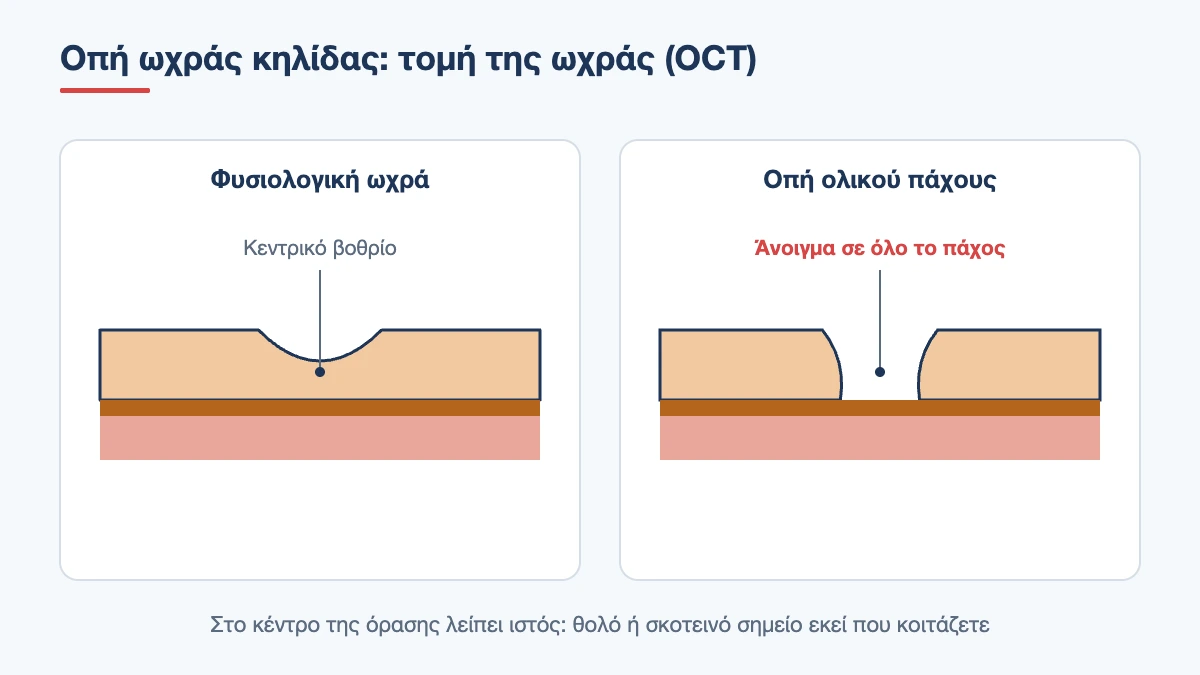
In a macular hole, the tissue at exactly this point opens up. The photoreceptors are not destroyed at first but are pulled apart to the sides, just as a hole opens in a stretched piece of fabric. This explains two things: why the patient sees objects “narrowing” or being “sucked in” towards the centre, and why, if the hole is closed in time, vision can recover to a large extent.
The hole is usually very small, from 0.1 to 0.6 millimetres. Its size should not be misleading, however: because it lies right at the centre, even an opening of a quarter of a millimetre is enough to make reading very difficult.
How common is it and who does it affect?
It is estimated that about 3 in 1,000 people over the age of 55 will develop a macular hole. Women are affected two to three times more often than men, and the typical age of onset is between 60 and 75. If there is a hole in one eye, the chance of one developing in the other eye in the following years is about 10 to 15%, unless the vitreous in the second eye has already fully detached, in which case the risk becomes very small.
Book an appointment with the doctor
An examination and clear advice on your case at our practice in Neo Psychiko.
Causes: why does a macular hole open?
Traction from the vitreous
The vast majority of holes are idiopathic, in other words they are not caused by another eye condition but by the normal ageing of the vitreous. The vitreous is the clear gel that fills the eye and, in younger years, is attached to the retina. With age it shrinks and separates, in a process called posterior vitreous detachment. In most people it separates smoothly. In some, however, it remains firmly attached right at the centre of the macula while it has come away all around. Every eye movement then transmits traction to the fovea, until the tissue gives way and opens.
Other causes
- High myopia: the elongated myopic eye has a thinner, more stretched retina, and holes appear at a younger age
- Injury: a hard blow to the eye, for example from a ball or a champagne cork, can cause a hole even in children and young people. Traumatic holes sometimes close on their own
- Chronic macular oedema due to diabetes, vein occlusion or inflammation
- Epiretinal membrane pulling tangentially on the macula
- Retinal detachment or previous surgery for it
A macular hole is not caused by reading, screens, tiredness or lifting heavy objects, and there is nothing you could have done to prevent it.
Symptoms
Symptoms usually develop over weeks and affect only central vision:
- blurred central vision that is not corrected with glasses
- distortion: straight lines bend or “break” in the middle, the letters of a word seem squashed together
- a feeling that a letter or part of the face you are looking at is missing
- a dark or grey spot exactly where you are focusing, in the more advanced stages
- difficulty reading, threading a needle or recognising faces at a distance
Peripheral vision remains normal, so the patient moves around comfortably. There is no pain or redness. Because the other eye compensates, many people notice the problem by chance, when they close their healthy eye, and by then the hole may already have been present for several months. A simple way to check is to look at a text or an Amsler grid with each eye separately from time to time.
A macular hole does not by itself cause flashes or a shower of floaters. If such symptoms or a shadow from the side appear, the periphery needs to be checked for a tear.
Stages and classification
A hole does not form from one moment to the next. It goes through stages that are clearly visible on OCT and determine treatment.
| Stage | What we see on OCT | Vision | Management |
|---|---|---|---|
| Impending hole | The vitreous pulls on the fovea, a cyst forms, there is no full-thickness opening | Slightly blurred, mild distortion | Monitoring, about half resolve on their own |
| Small full-thickness hole | Opening up to 0.25 mm | Moderately reduced | Surgery, excellent prognosis |
| Medium hole | Opening 0.25 to 0.4 mm | Clearly reduced | Surgery |
| Large hole | Opening over 0.4 mm | Very reduced, central scotoma | Surgery, often with special techniques |
As well as size, what matters is whether the vitreous is still attached to the edges of the hole and how long the hole has been present. Small, recent holes almost always close and give the best vision.
Pseudohole and lamellar hole: not the same thing
These are two terms that often confuse people reading their OCT report. A pseudohole is not a hole: it is an epiretinal membrane that wrinkles the macula and gives the impression of a hole, while the tissue at the centre is intact. A lamellar hole is a partial defect, where only the inner layers are missing and the outer layers, with the photoreceptors, remain in place. Both usually have much better vision than a true hole, progress slowly and in most cases are simply monitored.
Diagnosis
On slit lamp examination with the pupils dilated, the hole looks like a round, well-defined reddish spot at the centre of the macula. A classic clinical test is to project a thin line of light onto the macula: a patient with a true hole sees the line broken or narrowed in the middle.
The test that confirms the diagnosis, however, is optical coherence tomography (OCT). Within a few seconds and without any discomfort, it shows in cross-section whether the defect is full-thickness or partial, measures its diameter, and reveals whether the vitreous is still pulling on the edges and whether there is a membrane. With OCT we always check the other eye too, so that we know whether there is traction there that needs monitoring.
Treatment
When we wait
At the impending hole stage, where there is not yet a full-thickness opening, about half of cases resolve spontaneously once the vitreous releases the fovea. Here the right choice is a repeat OCT every few weeks. The same applies to lamellar holes and pseudoholes with good vision.
In contrast, a true full-thickness hole rarely closes on its own. Without treatment it usually enlarges over months and vision settles at a low level. There are no drops, medicines or laser treatments that close the hole. An intravitreal enzyme injection that was used for very small holes with traction has essentially been abandoned because of its low effectiveness.
The operation: vitrectomy with gas
The established treatment is vitrectomy. It is performed under local anaesthetic, takes about 45 minutes to an hour, and the patient goes home the same day. There are three steps:
- Removal of the vitreous through three tiny openings in the white of the eye, to eliminate any forward traction
- Peeling of the internal limiting membrane around the hole. This is the thinnest innermost layer of the retina. Removing it makes the tissue more elastic, so that the edges of the hole can come together
- Filling the eye with gas. The bubble gently presses on the edges of the hole and keeps them dry until they seal. The gas is absorbed on its own and replaced by the eye’s natural fluid
For very large or long-standing holes, variations of the technique are used, such as an inverted flap of internal limiting membrane placed over the hole, which acts as a bridge for healing.
Head positioning after surgery
This is the part that worries patients most. Because the bubble always rises upwards, the face must point downwards for it to rest against the macula, which is at the back of the eye. In the past, strict face-down positioning was required for one to two weeks. Today, with modern gases and internal limiting membrane peeling, a few days or simply avoiding lying on your back is often enough for small holes, while for larger holes positioning is usually kept up for 3 to 7 days. The exact plan is set by the surgeon according to the size of the hole.
Special pillows and chairs can be rented to make this period more bearable. It helps to have your home organised in advance: a table to rest your forehead on, audiobooks or the radio, and someone to help you during the first few days.
While there is gas in the eye
- Vision is very blurred. As the bubble shrinks, you see a horizontal line that wobbles and moves down day by day, like a spirit level
- Flying is not allowed, nor is travelling to high altitude, because the gas expands and the eye pressure can rise dangerously
- If you need a general anaesthetic for any reason, tell the anaesthetist that you have gas in your eye, because certain anaesthetic gases are contraindicated
- Depending on the type of gas, absorption takes 2 to 8 weeks
Results: how much will vision improve?
The hole closes after the first operation in more than 9 out of 10 patients, and in almost all when it is small and recent. Closure is confirmed with OCT, often within the first few weeks.
Anatomical closure and vision are not the same thing, however. The photoreceptors need months to reorganise, and vision continues to improve for 6 to 12 months, sometimes longer. About 7 to 8 out of 10 patients gain at least two lines on the chart, and most can read again with that eye. Distortion is greatly reduced, but a small residual amount is common.
Three factors predict a better outcome: a small hole diameter, better vision before surgery and, above all, a short duration of symptoms. Holes operated on within the first six months do clearly better than those that have been present for years. This is why we do not recommend waiting once the diagnosis has been made.
Book an appointment with the doctor
An examination and clear advice on your case at our practice in Neo Psychiko.
Risks of surgery
- Cataract: almost all patients who still have their natural lens will develop a cataract within 1 to 2 years. This is why the two operations are combined in many patients
- Retinal tear or detachment: about 1 to 2 in 100 operations
- Failure to close or reopening: in a small percentage, mainly in large and long-standing holes. A second operation can be performed
- Raised eye pressure in the first few days, which is treated with eye drops
- Intraocular infection (endophthalmitis): very rare, but serious
Contact your ophthalmologist immediately if, after surgery, you have severe pain that does not ease with painkillers, nausea, a sudden worsening of vision or a shadow rising from the periphery.
What happens if it is not operated on?
A full-thickness hole that is not treated usually enlarges, and central vision drops to a level where only the largest letters on the chart can be read. The eye does not go blind, because peripheral vision is preserved, and the patient remains independent, especially if the other eye sees well. The decision to operate is ultimately a personal one. It is worth making it, however, knowing that time matters, that the chance of success is high and that the other eye also carries a small risk in the future.
Macular hole and high myopia
In eyes with more than 6 dioptres of myopia, macular holes have particular features. They appear earlier, often before the age of 50, and can coexist with splitting of the retinal layers, a condition called myopic foveoschisis, or with a posterior staphyloma, an outward bulging of the back wall of the eye. Most importantly, in a myopic eye a macular hole can lead to a retinal detachment that starts from the centre, something that is extremely rare in non-myopic eyes. For this reason, people with high myopia benefit from periodic OCT checks even without symptoms, and surgery in these patients is planned with greater care, because closure rates are somewhat lower.
Daily life after recovery
Once the gas has been absorbed and OCT has confirmed that the hole has closed, there are no permanent restrictions. You can exercise, lift weights, swim and travel as before. A new glasses prescription is usually needed after 2 to 3 months, because the eye’s refraction changes, especially if cataract surgery was performed at the same time. During the period when the vision of the two eyes differs a lot, quite a few patients find it difficult to judge distances, for example when going down stairs or pouring water into a glass. This improves as the operated eye recovers and the brain adapts. Follow-up visits are usually on the first day, at one week, at one month and then at 3 and 6 months, with OCT at each visit.
Preparing for surgery: a practical guide
- Arrange for someone to accompany you on the day of surgery and to stay with you the first night
- Tell us about all your medicines, especially anticoagulants. Do not stop anything without instructions
- If face-down positioning is needed, try out the pillow arrangement in advance and decide where you will sleep
- Postpone air travel for at least two months and do not book tickets before the surgeon tells you the gas has been absorbed
- Do not drive while there is gas in the eye
- Plan to take 2 to 4 weeks off work, depending on your job
When to have an examination
If lines look broken with one eye, if letters are missing from words or if there is a blurred spot exactly where you are looking, book an appointment within a few days. With OCT the diagnosis is made at the same visit, and we know immediately whether it is a hole, a membrane or something else, as well as how soon we need to act. Practice telephone 210 677 0007 or use the contact form.
A blurred spot in the centre of your vision?
Early diagnosis with OCT improves the outcome of treatment. Book an appointment within a few days.
Frequently Asked Questions about Macular Hole
Can a macular hole close on its own?
A true full-thickness hole rarely closes spontaneously, only in a few cases of very small or traumatic holes. The early stage, where the vitreous simply pulls on the fovea without a full opening, resolves on its own in about half of cases. OCT shows which category you are in.
Is a macular hole the same as macular degeneration?
No. A macular hole is a mechanical problem, an opening caused by traction from the vitreous, and it is corrected surgically. Age-related macular degeneration is wear of the deeper layers of the macula and is managed with monitoring or intravitreal injections. The symptoms are similar, which is why OCT is needed.
How many days do I need to stay face down?
It depends on the size of the hole and the technique. For small holes, a few days or simply avoiding sleeping on your back is often enough. For larger ones, 3 to 7 days are usually recommended. Your surgeon will give you specific instructions before the operation.
When can I fly after macular hole surgery?
Only once the gas has been fully absorbed, which takes 2 to 8 weeks depending on its type. At altitude the bubble expands and can raise the eye pressure dangerously. Always wait for confirmation from your ophthalmologist.
Will my vision fully recover?
Most patients improve significantly and can read again with the operated eye, but vision rarely becomes exactly the same as before. Improvement continues for 6 to 12 months. The result is better when the hole is small and operated on early.
Is there a risk of a macular hole in the other eye?
Yes, but it is small, about 10 to 15% over the years. If the vitreous in the other eye has already fully detached, the risk is minimal. This is why both eyes are checked with OCT at every visit.
What happens if I do not have the operation?
The hole usually enlarges and the central vision of the eye drops significantly, while peripheral vision is preserved. The eye does not go completely blind. However, as time passes, the benefit that later surgery can offer decreases.
Medically reviewed by: Dr. Vasileios Skouteris MD, FEBO