
Written by Vasileios Skouteris MD, FEBO, Ophthalmic Surgeon
Page contents
- What is ptosis?
- Ptosis or simply “heavy” eyelids?
- Types and causes
- Symptoms
- When is a droopy eyelid an emergency?
- Diagnosis
- Treatment
- Ptosis in children: when is surgery needed?
- Recovery
- Risks and realistic expectations
- What you can do to help prevent it
- When to book an appointment
- Frequently Asked Questions about Ptosis
Ptosis, or a droopy eyelid, is when the upper eyelid sits lower than normal, so that it covers part of the pupil and makes the eye look half-closed. It can be present from birth or develop later in life, usually because the tendon that lifts the eyelid has loosened. It is not just a cosmetic issue: it limits your field of vision, causes fatigue and, when it appears suddenly, can be the first sign of a neurological problem. Treatment is, as a rule, surgical.
What is ptosis?
In its normal position, the margin of the upper eyelid covers just 1 to 2 millimetres of the top of the iris and leaves the whole pupil clear. The eyelid is raised mainly by the levator muscle, which starts deep in the eye socket and ends in a thin, broad tendon, the aponeurosis, attached to the tarsal plate of the eyelid. A smaller role is played by Müller’s muscle, which provides the last 1 to 2 millimetres of lift and is controlled by the sympathetic nervous system. That is why our eyes open wider when we are alert and feel “heavy” when we are sleepy.
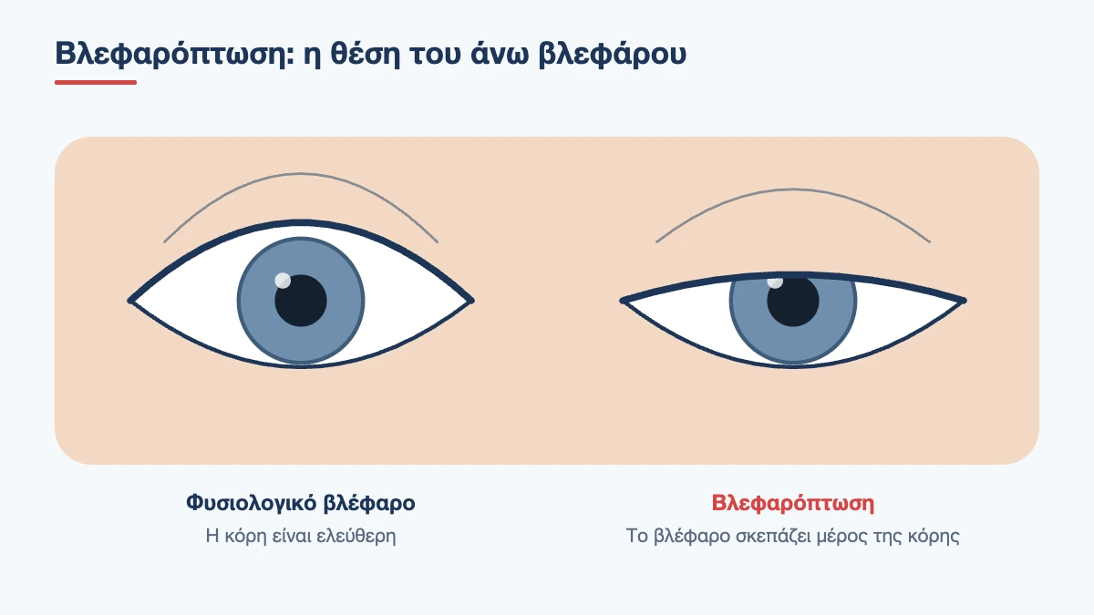
When any link in this chain fails, whether the muscle, the tendon, the nerve or the junction between nerve and muscle, the eyelid drops. The droop may be minimal, barely visible in photographs, or so severe that the eyelid covers the entire pupil. It may affect one eye or both, often to a different degree.
Ptosis or simply “heavy” eyelids?
This is the most common source of confusion and it matters in practice, because the two conditions are corrected with different operations.
- Dermatochalasis: there is excess skin on the upper eyelid that hangs like a curtain over the lashes. The eyelid margin itself is in the correct position. It is corrected with blepharoplasty, that is, removal of the excess skin
- Brow ptosis: the eyebrow has descended and pushes the skin down. The eyelid is not the cause
- True ptosis: the eyelid margin itself, where the lashes grow, sits low. It can only be corrected by strengthening the lifting mechanism
All three conditions very often occur together in the same person. If only a blepharoplasty is performed on an eyelid that also has ptosis, the eye will stay half-closed and the patient will be disappointed. That is why accurate measurement before any eyelid surgery is essential. A false appearance of ptosis can also be caused by an eye that has sunk back into the socket, or by an eyelid that only looks low because the other one is abnormally wide open, as happens in thyroid eye disease.
Book an appointment with the doctor
An examination and clear advice on your case at our practice in Neo Psychiko.
Types and causes
Congenital ptosis
This is present from birth and is usually caused by incomplete development of the levator muscle, in which fibrous and fatty tissue takes the place of muscle fibres. This muscle neither contracts nor relaxes properly: the eyelid is low when the child looks straight ahead, but stays higher than normal when the child looks down. It usually affects one eye and is not linked to any other health problem. Children often lift their chin or wrinkle their forehead to see under the eyelid.
The main risk in children is amblyopia (lazy eye). If the eyelid covers the pupil, the eye does not receive an image and does not develop vision. Even when the pupil is clear, the weight of the eyelid on the cornea often causes astigmatism, which in turn can lead to amblyopia. Every child with a droopy eyelid needs an early paediatric eye examination and regular follow-up.
Aponeurotic (age-related) ptosis
This is by far the most common form in adults. The muscle is healthy and strong, but its tendon has thinned, stretched or come away from the tarsal plate of the eyelid. Apart from age, it can be caused or accelerated by:
- many years of contact lens wear, especially rigid lenses, because of the daily pulling on the eyelid
- chronic eye rubbing, for example with allergies
- previous eye surgery, such as cataract surgery, where an eyelid speculum is used
- repeated swelling of the eyelids
Its typical features are that the eyelid crease rises higher, the eyelid looks thin and “hollow”, and the droop worsens towards the evening or when looking down, and therefore when reading.
Neurogenic ptosis
Here the problem lies in the nerve that sends the signal. Two conditions are particularly important:
- Third nerve (oculomotor) palsy: marked ptosis, an eye that turns outwards and downwards, and double vision. If the pupil is dilated and there is a headache, it may be caused by an aneurysm pressing on the nerve, which is an emergency
- Horner syndrome: mild ptosis of 1 to 2 millimetres, with a smaller pupil on the same side. It is caused by damage to the sympathetic pathway, which runs through the chest and neck. When it comes with pain in the neck or face, carotid artery dissection must be ruled out urgently
Myogenic ptosis
This is caused by a disease of the muscle itself or of its connection with the nerve. The best known is myasthenia gravis, in which the droop changes during the day: it is mild or absent in the morning and worsens with fatigue, often together with double vision. In many patients the eyes are the first or only sign of the disease. Rarer causes are mitochondrial myopathies and muscular dystrophies, in which the ptosis affects both sides, is symmetrical and progresses slowly over years.
Mechanical and traumatic ptosis
The eyelid can drop because something is weighing it down, such as a large chalazion, a tumour or swelling, or because a scar is holding it. An injury that cuts the levator muscle or trauma to the eye socket has the same effect. Finally, temporary ptosis lasting a few weeks can occur after botulinum toxin injections in the forehead, when the drug spreads towards the levator muscle. It resolves completely on its own.
Symptoms
- one or both eyes look smaller, sleepy or asymmetrical
- loss of the upper part of the field of vision, noticeable when driving, on stairs or when looking at high shelves
- difficulty reading, because the eyelid drops further when you look down
- fatigue and forehead ache towards the end of the day, from the constant effort to raise the eyebrows
- deep horizontal forehead lines and permanently raised eyebrows
- a need to tilt the head back or lift the eyelid with a finger
- comments from people around you that you look tired when you are not
When is a droopy eyelid an emergency?
A slow, gradual droop over years is almost always benign. On the other hand, you need a same-day examination, and often a neurological assessment and imaging, if the eyelid has dropped:
- suddenly, within hours or a few days
- together with double vision
- together with pupils of unequal size
- together with a severe headache or pain in the neck, face or behind the eye
- together with difficulty swallowing, speaking or breathing, or weakness in the arms or legs
- after an injury
The same applies to babies whose eyelid covers the pupil: here the urgency is not neurological but concerns the development of vision.
Diagnosis
The examination is clinical and is based on a few simple but precise measurements, which also determine the type of surgery:
- Eyelid position: we measure in millimetres the distance from the upper eyelid margin to the centre of the pupil. Normally it is 4 to 5 millimetres. Below 2, the ptosis is considered significant
- Levator function: how far the eyelid travels from looking down to looking up, with the eyebrow held still. Over 12 millimetres is normal, under 4 is poor. This is the most important measurement for choosing the technique
- Height of the eyelid crease and position of the eyebrow
- Pupils and eye movements, to rule out a neurological cause
- Fatigue testing: sustained upward gaze for one minute, or an ice pack on the eyelid for two minutes. If the ptosis worsens with fatigue or improves with cold, myasthenia gravis is suspected
- Phenylephrine drop test: if the eyelid rises within a few minutes, the patient is a candidate for surgery from the inner side of the eyelid
- Ocular surface: we check the tears and how well the eye closes, because underlying dry eye affects how far the eyelid can safely be lifted
Visual field tests, with the eyelid in its natural position and then lifted with tape, objectively document how much field of vision is gained. When myasthenia gravis is suspected, blood tests for specific antibodies and a neurological assessment are requested, while neurogenic ptosis requires MR or CT angiography. It is helpful to bring old photographs: they show when the ptosis began and how quickly it is progressing.
Treatment
When the ptosis is mild and does not bother you, nothing needs to be done. When it is caused by another disease, such as myasthenia gravis or a nerve palsy, the cause is treated first and the eyelid often recovers. In all other cases correction is surgical. There are three main techniques and the choice depends mainly on the strength of the levator muscle.
Levator aponeurosis advancement
This is the most common operation in adults. Through an incision hidden in the natural crease of the eyelid, the surgeon finds the loosened tendon, shortens it and reattaches it to the tarsal plate. It is performed under local anaesthesia with the patient awake, which is an advantage: the patient is asked to open their eyes and sit up, so that the height and curve of the eyelid can be adjusted on the spot. If there is also excess skin, it is removed through the same incision, so the ptosis correction is combined with blepharoplasty.
Posterior (internal) approach
In mild ptosis with a good response to the phenylephrine drop test, Müller’s muscle can be shortened together with the conjunctiva from the inner surface of the eyelid. There is no skin incision, the operation is short and the result is predictable.
Frontalis sling
When the levator muscle is very weak, as in most severe congenital ptosis, there is no point in shortening it. Instead, the eyelid is connected to the forehead muscle with a thin silicone band or with fascia taken from the patient, so that it rises when the eyebrow is raised. In children it is performed under general anaesthesia.
Is there a non-surgical option?
For those who cannot have or do not want surgery, there are special spectacle frames with a thin support (ptosis crutch) that holds the eyelid up. They are mainly useful in neurogenic or myogenic ptosis. In some countries there are also eye drops that stimulate Müller’s muscle and raise the eyelid by about one millimetre for a few hours. These are a temporary aid in mild cases, not a treatment. Exercises, massage and creams do not correct true ptosis.
Book an appointment with the doctor
An examination and clear advice on your case at our practice in Neo Psychiko.
Ptosis in children: when is surgery needed?
If the eyelid covers the pupil or has already caused amblyopia, surgery is performed early, even in infancy. If the pupil is clear and vision is developing normally, it is preferable to wait until the age of 3 to 5, shortly before school, when measurements are more reliable and the tissues more mature. In the meantime the child is monitored with vision and refraction checks every few months, and glasses or patching are given if needed. Parents should know that after surgery the eye may stay slightly open during sleep and that, as the child grows, a second correction may be needed.
Recovery
After surgery, cold compresses are applied for the first 48 hours and sleeping with your head raised is recommended. Swelling and bruising peak on the second to third day and settle within 1 to 2 weeks. Stitches are removed after 5 to 7 days, unless they are absorbable. Most people return to office work within a week, while strenuous exercise and swimming are avoided for 2 to 3 weeks.
In the first few weeks the eye may not close completely and may become dry, so artificial tears are used during the day and a lubricating ointment at night. The final height and shape of the eyelid can be judged after about 2 to 3 months, once all the swelling has gone down.
Risks and realistic expectations
The aim is to clear the pupil, open up the field of vision and make both eyes look as symmetrical as possible. Perfect symmetry, to within half a millimetre, is not always achievable, and it is good to say this from the outset.
- Under-correction or over-correction: the eyelid stays slightly lower or rises slightly higher than intended. In a small percentage, about 5 to 10%, an additional correction is needed
- Asymmetry in height, crease or curve
- Incomplete closure and dryness, usually temporary
- Drooping of the other eyelid: when one side is corrected, the brain reduces its effort and reveals a mild ptosis on the other side, which was there but not visible
- Haematoma or infection: rare
- Recurrence over the years, as the tissues continue to loosen
What you can do to help prevent it
Age-related loosening cannot be fully prevented, but some habits protect the tendon: avoid rubbing your eyes hard, treat the allergies that make you rub them, remove contact lenses without pulling the upper eyelid and limit the hours you wear them where possible. If you notice that one eyelid is lower than in older photographs, an examination will show whether it is simple loosening or something that needs investigating.
When to book an appointment
If your eyelid feels heavy, limits your field of vision or has changed over recent months, an examination with precise measurements will clarify whether it is ptosis, excess skin or a combination, and which correction suits you. If the droop appeared suddenly or comes with double vision, headache or unequal pupils, ask to be seen the same day. Call the clinic in Neo Psychiko, Athens, on 210 677 0007 or use the contact form.
Does your eyelid feel heavy?
With precise measurements we can tell ptosis apart from loose skin. Book an appointment for an assessment.
Frequently Asked Questions about Ptosis
Can ptosis be corrected without surgery?
True ptosis cannot be permanently corrected with exercises, creams or massage. There are temporary aids, such as special supports fitted to glasses, which help people who cannot have surgery. When the ptosis is caused by another condition, such as myasthenia gravis, treating that condition may make it improve.
What is the difference between ptosis and blepharoplasty?
Ptosis is a condition: the eyelid margin sits low because the lifting mechanism has weakened. Blepharoplasty is an operation that removes excess skin. When ptosis and loose skin occur together, both corrections are done at the same time, through the same incision.
Is the surgery painful and how long does it take?
It is performed under local anaesthesia and takes about 45 to 60 minutes per eyelid. You feel no pain during the operation, only touch and a slight pulling sensation. Afterwards the discomfort is mild and is controlled with a simple painkiller and cold compresses.
How soon can I go back to work?
Most people return to office work within a week. Bruising and swelling settle within 1 to 2 weeks. Strenuous exercise, swimming and eyelid make-up should be avoided for 2 to 3 weeks.
Can ptosis come back after surgery?
Yes, over the years, because the tissues continue to loosen with age. In congenital ptosis operated on in childhood, a second correction later on is not unusual. If the ptosis recurs, the operation can be repeated.
My eyelid drooped after Botox. Is it permanent?
No. It happens when the drug spreads towards the muscle that lifts the eyelid, and it resolves completely within a few weeks as the effect of the toxin wears off. Your ophthalmologist can prescribe drops that temporarily lift the eyelid during this time.
Does ptosis affect my child’s vision?
It can. If the eyelid covers the pupil or causes astigmatism, the eye is at risk of becoming amblyopic (lazy eye). That is why vision and refraction checks are needed early, along with regular follow-up, even if surgery is planned for later.
Medically reviewed by: Dr. Vasileios Skouteris MD, FEBO