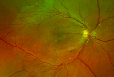
Diabetic macular oedema is one of the conditions that affect the macula, a term many people have heard of nowadays because of the changes that can occur in this area. But what is the macula? It is the anatomical part of the eye that gives us our sharpest vision. It lies at the centre of the retina and takes its name (macula lutea) from the yellowish appearance it shows when examined during fundoscopy, while the retina is the tissue of the eye that receives light and turns it into an image. It is easy to see, therefore, that the proper function of the retina, and above all of its central part, the macula, is essential for good vision. Unfortunately, there are several conditions that affect the macula. The best known are age-related macular degeneration, diabetic maculopathy and myopic maculopathy.
Diabetic maculopathy is the damage to the macula caused by the microangiopathy of diabetes. Diabetes mellitus, as we know, is a microangiopathy that mainly affects parts of the body with a large number of small blood vessels called capillaries. Our eyes are one of these parts of the body, together with the kidneys and the nerves, that are made up of a large number of capillaries and depend on them to function properly. High blood sugar levels cause micro-injuries to the capillaries, resulting in changes such as small haemorrhages and small leaks. These leaks lead to fluid building up in the macula, which we call diabetic macular oedema.
What are the effects of this oedema?
First of all, in terms of vision, there is progressive blurring, mainly of the central visual field. The larger the oedema, the greater the disturbance to vision. In addition, a condition called metamorphopsia often develops. In metamorphopsia, objects look distorted. This is particularly noticeable with objects that have straight lines, such as doors and windows, which look wavy. The symptoms usually progress slowly, but there are cases of rapid deterioration.
In recent years, diagnosis has become much simpler with the use of a machine called optical coherence tomography (OCT). With optical coherence tomography (OCT) we obtain, in a very short time and without radiation or pain, cross-sectional images of the macula which, together with fundoscopy, give us all the information we need to make a reliable diagnosis and move on to effective treatment.
Finally, it is worth stressing how much the results of treatment have improved, provided, of course, that the treating doctor has the necessary specialist expertise and the patient cooperates well. With a choice between treatment with intravitreal injections and two different types of laser, we are in a position to achieve very good results. Intravitreal injections (injections into the eye) are not painful, and you do not see anything during the injection. All you feel is slight pressure at the edge of the eye, which is why the great majority of patients keep up with their treatment and have good results. In some other cases we choose laser treatment, which has been tried and tested for many years and offers important solutions.
In conclusion, correct and prompt treatment is of the utmost importance in managing this increasingly frequently diagnosed condition.
Questions about your case?
Dr. Skouteris will examine you at his practice in Neo Psychiko and explain your options.

Ophthalmic surgeon trained at the John Radcliffe University Hospital in Oxford and Fellow of the European Board of Ophthalmology (FEBO).
Full profileRelated articles
Aftercare Instructions after an Intravitreal Injection
Please follow the aftercare instructions below after your intravitreal injection to avoid a possible infection or other serious complication from the injection:…
Read the article
Protecting Your Eyes in Summer: The 3 Most Common Mistakes
Proper summer eye protection from solar radiation is extremely important. It is widely known how beneficial the sun can be for our…
Read the article
Eye Floaters: How Are They Linked to a Retinal Tear?
Eye floaters are usually harmless, but they can sometimes be linked to a retinal tear. A retinal tear is a common condition,…
Read the article