
Written by Vasileios Skouteris MD, FEBO, Ophthalmic Surgeon
Page contents
- What is an epiretinal membrane?
- How common is it?
- Causes: why does it form?
- Symptoms
- What can it be confused with?
- Diagnosis
- Stages and progression
- Treatment
- What results to expect
- Risks and complications
- Epiretinal membrane and cataract surgery
- Questions worth asking before you decide
- Living with a membrane under monitoring
- When to have an examination
- Frequently Asked Questions about Epiretinal Membrane
An epiretinal membrane is a very thin, transparent layer of fibrous tissue that grows on the surface of the macula, the centre of the retina. When the membrane contracts, it pulls and wrinkles the retina beneath it, resulting in blurred and distorted central vision. It is common after the age of 60, is diagnosed with OCT and, when it causes problems, is removed surgically.
What is an epiretinal membrane?
The retina is the light-sensitive layer that lines the inside of the eye, rather like the film in an old camera. At its centre lies the macula, an area a few millimetres across that is responsible for sharp, detailed vision: reading, recognising faces, driving. To work properly, the macula must be completely smooth and flat.
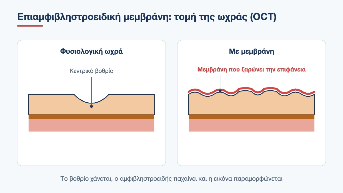
An epiretinal membrane forms when cells migrate and multiply on this surface, creating a sheet of tissue that looks like transparent cling film. At first it is so thin that it affects nothing. Over time, however, it can thicken and contract, just as a scar on the skin contracts. The retina beneath then wrinkles, thickens and loses its normal central dip.
In the medical literature and in test reports you will come across it under many names: epimacular membrane, macular pucker, cellophane maculopathy, or the abbreviation ERM. They all describe the same condition at different levels of severity.
How common is it?
It is one of the most common findings at the macula. Some degree of membrane is found in about 1 in 10 people over the age of 60, and the proportion rises with age. Most people will never know unless they have an OCT scan, because their membrane is thin and causes no symptoms. In 2 to 3 out of 10 patients it affects both eyes, usually not to the same degree.
Book an appointment with the doctor
An examination and clear advice on your case at our practice in Neo Psychiko.
Causes: why does it form?
Idiopathic membrane
In most cases there is no other condition in the eye and the membrane is called idiopathic. The main cause is the normal ageing of the vitreous, the gel that fills the inside of the eye. With age the vitreous becomes more liquid and separates from the retina, a process called posterior vitreous detachment. As it separates, it can leave remnants behind or cause microscopic breaks in the inner surface of the retina, through which cells emerge and spread over the macula.
Secondary membrane
In other patients the membrane is the result of another condition that caused inflammation or healing inside the eye:
- a retinal tear or retinal detachment, as well as their treatment with laser or cryotherapy
- diabetic retinopathy
- retinal vein occlusion
- intraocular inflammation (uveitis)
- eye injury
- previous intraocular surgery
Secondary membranes also occur at younger ages and are often thicker. Their prognosis depends mainly on the underlying condition.
Risk factors
The most important factor is age. This is followed by diabetes, a membrane in the other eye and a history of surgery or inflammation. There is no proven link with eye strain, reading, screens or diet, and there is nothing the patient could have done to avoid it.
Symptoms
The picture varies a great deal. A thin membrane may be a chance finding at a routine check-up. When it starts to pull on the macula, however, characteristic symptoms appear:
- Distorted images (metamorphopsia): straight lines, such as tiles, window frames or lines of text, look wavy or broken. This is the most typical symptom
- Blurred central vision: difficulty reading small print that is not corrected by changing glasses
- Different image size: objects look larger or smaller with the affected eye than with the other, which makes it harder for the two eyes to work together
- Double vision in one eye or a feeling that letters “overlap each other”
- Tiredness when reading and a need to close one eye
Peripheral vision is not affected. An epiretinal membrane does not cause pain, redness or watering, and does not lead to complete blindness. Because the other eye often compensates for the problem, many people discover it by chance, when they close their good eye.
How to check for distortion yourself
The Amsler grid is a simple sheet of squared paper with a dot in the centre. You look at it with each eye separately, wearing your reading glasses, at reading distance. If the lines around the dot look crooked, are missing or are blurred, there is something at the macula that needs checking. The same test is used by patients with macular degeneration, and it is a useful way to monitor whether anything changes between visits.
What can it be confused with?
Distorted central vision is not unique to an epiretinal membrane. The same symptom can be caused by:
- the wet form of age-related macular degeneration, which, however, progresses quickly and needs prompt treatment with injections
- a macular hole, where tissue is missing at the centre and a dark spot appears
- vitreomacular traction, where the vitreous remains stuck to the centre and pulls it forwards
- macular oedema due to diabetes or vein occlusion
- central serous chorioretinopathy, usually in younger patients
These conditions have completely different treatments and different levels of urgency. This is why any new distortion needs an examination within a few days, not a wait of months.
Diagnosis
Clinical examination
After measuring your vision, the pupils are dilated with drops and the fundus is examined with the slit lamp. The membrane appears as a glistening reflection on the macula, and in more advanced forms the folds of the retina and the distortion of the small blood vessels can be seen. The periphery is also checked carefully, to rule out a tear that could be the cause.
Optical coherence tomography (OCT)
OCT is the test that makes the diagnosis and determines management. It is painless, takes a few minutes and shows the layers of the retina in cross-section, with micrometre precision. On OCT we see the membrane as a white line on the surface, measure the thickness of the macula, and check whether the central foveal pit is preserved and whether the inner or outer layers have been disrupted. These findings allow staging and, above all, give an estimate of how much vision may recover after surgery.
Comparing scans over time is just as useful. A membrane that remains unchanged for two years is managed differently from one that thickens from one six-monthly check to the next.
Other tests
When a vascular condition or degeneration is suspected, OCT angiography or fluorescein angiography may be requested. Fundus photography is used for documentation and comparison.
Stages and progression
In the early stages the membrane is thin, the foveal pit is preserved and vision is normal or almost normal. In the intermediate stages the pit flattens, the macula thickens and distortion appears. In the advanced stages the retinal layers become disorganised and vision drops significantly.
The reassuring point is that most membranes do not follow this whole course. In long-term follow-up, the majority remain stable, some worsen slowly and a small percentage resolve on their own, when the membrane spontaneously separates from the macula. This is why the diagnosis does not automatically mean surgery.
Treatment
Monitoring
When vision is good and the distortion does not interfere with daily life, the right choice is monitoring with OCT, usually every 6 to 12 months, and self-checking with the Amsler grid at home. There are no drops, tablets, dietary supplements or exercises that dissolve the membrane or stop it from progressing. Changing glasses does not solve the problem either, although a correct prescription helps you make the most of the vision you have.
When is surgery recommended?
The decision is not based only on the OCT image, but mainly on how much the patient is troubled. We discuss surgery when:
- vision has dropped to a level that makes reading, driving or work difficult
- the distortion is marked and bothersome, even if visual acuity remains relatively good
- OCT shows progressive worsening from one examination to the next
Timing matters. Results are better when surgery is performed before vision drops too much and before the photoreceptor layers are permanently disrupted. Waiting “until I can’t see at all” is not a good strategy. On the other hand, this is not emergency surgery: there are weeks or even months available to plan it properly.
Vitrectomy with membrane peeling
The operation is called a vitrectomy. It is usually performed under local anaesthetic with light sedation, takes about 30 to 60 minutes and the patient goes home the same day. Through three tiny openings less than half a millimetre in diameter, which as a rule do not need stitches, the surgeon removes the vitreous and then, using fine forceps under high magnification, grasps the edge of the membrane and peels it off the macula. A special dye is often used to make the tissue more visible, and the internal limiting membrane, the thinnest innermost layer of the retina, is often removed as well to reduce the chance of recurrence.
In patients who already have an early cataract, it is often removed during the same operation, because vitrectomy speeds up clouding of the lens in any case.
After surgery
The eye is covered for one day and eye drops are used for 3 to 4 weeks. In the first few days a foreign body sensation, redness and blurred vision are normal. If an air or gas bubble has been placed, vision will be very blurred until it is absorbed, and during this time flying or travelling to high altitude is not allowed. Most people return to office work within 1 to 2 weeks, while heavy physical work and swimming are avoided for about a month.
What results to expect
Visual recovery is slow, and this is the first thing anyone having surgery needs to know. The retina needs time to unfold: improvement continues for 6 to 12 months. About 7 to 8 in 10 patients gain two or more lines on the vision chart, and most report a clear reduction in distortion.
The second thing that must be clear is that vision rarely returns to completely normal. A small degree of distortion usually remains, and the final result depends on how long the membrane had been present, how good vision was beforehand and the condition of the outer layers on OCT. The realistic aim of surgery is to stop the deterioration and substantially improve functional vision, not to “make the eye as it was at 30”.
Book an appointment with the doctor
An examination and clear advice on your case at our practice in Neo Psychiko.
Risks and complications
Modern vitrectomy is a safe operation, but like any surgery it has risks that should be discussed before the decision is made:
- Cataract: this is the most common consequence. In most patients who have not already had cataract surgery, the lens becomes cloudy within the next 1 to 2 years
- Retinal tear or detachment: about 1 to 2 in 100 operations. It can be treated but requires further surgery
- Intraocular infection (endophthalmitis): very rare, less than 1 in 1,000, but serious
- Recurrence of the membrane: in a small percentage, even lower when the internal limiting membrane is also removed
- A temporary rise in eye pressure or a small haemorrhage, which usually settle with treatment
After surgery, a sudden drop in vision, severe pain, flashes or a curtain-like shadow in your field of vision are reasons to contact your ophthalmologist immediately.
Epiretinal membrane and cataract surgery
Many patients find out they have a membrane during the pre-operative assessment for cataract, and this is one of the reasons why an OCT of the macula is worth doing before any cataract operation. Having a membrane does not rule out surgery, but it affects two decisions. The first concerns the type of intraocular lens: multifocal lenses require a healthy macula to perform well, so in an eye with a membrane a monofocal or extended depth of focus lens is usually preferred. The second concerns expectations: if part of the blurred vision is due to the membrane, removing the cataract will help, but it will not eliminate the distortion.
After cataract surgery, macular oedema is slightly more common in eyes with a membrane, which is why anti-inflammatory drops are often given for longer and an OCT check is carried out in the first month.
Questions worth asking before you decide
Vitrectomy is a planned decision and you have time to find out what you need to know. When talking with your ophthalmologist or retinal surgeon, it is helpful to ask:
- What stage is my membrane at, and has it changed since the previous OCT?
- How much of the drop in vision is due to the membrane and how much to cataract or another cause?
- What improvement is realistic in my case, based on the outer layers of the macula?
- Will the cataract be removed at the same time?
- Will gas be needed and, if so, for how many days will I be unable to fly?
- What will happen if I choose to wait another six months?
The answers help you weigh the benefit against the risk based on your own needs. Someone who reads and drives every day has a different threshold from someone who is not troubled in their activities.
Living with a membrane under monitoring
For the many patients who do not need surgery, daily life does not change. You can read, work on the computer, exercise and travel without restrictions, because none of these makes the membrane worse. Good lighting for reading, an up-to-date glasses prescription and checking with the Amsler grid once a week all help. If you have diabetes, good blood sugar control protects the retina as a whole. And because a membrane can also appear in the other eye, regular check-ups always cover both eyes.
When to have an examination
Book an appointment if straight lines look wavy to you, if letters are distorted or if one eye sees objects at a different size from the other. If the distortion appeared suddenly, over a few days, do not put it off. At the practice in Neo Psychiko the diagnosis is made at the same visit with OCT, and we discuss whether monitoring is enough or whether the time has come for surgery. Telephone 210 677 0007 or use the contact form.
Do straight lines look wavy to you?
With OCT the diagnosis is made at the same visit. Book an appointment for a macular examination.
Frequently Asked Questions about Epiretinal Membrane
Is an epiretinal membrane dangerous?
It does not threaten the eye and does not lead to blindness. It affects only central vision, while peripheral vision remains intact. In most patients it stays stable for years. However, when it causes significant blurring or distortion, delaying surgery may limit how much vision recovers.
Are there eye drops or medicines that dissolve the membrane?
No. No medication, supplement or exercise removes or shrinks an epiretinal membrane. The only options are monitoring, when symptoms are mild, and surgical removal with vitrectomy, when vision is significantly affected.
When should an epiretinal membrane be operated on?
When blurred or distorted vision makes everyday activities such as reading and driving difficult, or when OCT shows steady worsening. It is not an emergency operation, but results are better when it is performed before vision drops too much.
How long is the recovery after vitrectomy?
A return to light activities is possible within 1 to 2 weeks and eye drops continue for about a month. Vision, however, improves gradually over 6 to 12 months, as the retina slowly returns to its normal shape.
Will I see perfectly after surgery?
Most patients see noticeably better and with less distortion, but vision rarely becomes completely normal. The final result depends on how long the condition has been present, vision before surgery and the condition of the macular layers on OCT.
Can the membrane come back?
Recurrence is uncommon, especially when the internal limiting membrane is also removed during surgery. If it does happen and causes symptoms, the membrane can be removed again. This is why an OCT check is also recommended after surgery.
Is an epiretinal membrane related to macular degeneration?
No, they are different conditions. The membrane lies on the surface of the retina and is a mechanical problem, while macular degeneration affects the deeper layers beneath it. They can coexist in the same eye, and OCT clearly distinguishes between them.
Medically reviewed by: Dr. Vasileios Skouteris MD, FEBO