
Written by Vasileios Skouteris MD, FEBO, Ophthalmic Surgeon
Page contents
- What is the vitreous body?
- How and why does the vitreous detach?
- Vitreous detachment and retinal detachment are not the same
- How common is it and who gets it earlier?
- Symptoms
- When is it an emergency?
- Diagnosis
- Possible complications
- Treatment
- What to do and what to avoid in the first few weeks
- Vitreous detachment after cataract or refractive surgery
- What to mention when you call for an appointment
- The psychological side: when floaters cause anxiety
- Flashes from the eye or from migraine?
- Will it happen in the other eye?
- When to book an appointment
- Frequently Asked Questions about Posterior Vitreous Detachment
Posterior vitreous detachment is the separation of the gel that fills the eye from the retina. It is a natural part of ageing, happens to most people after the age of 60 and shows up as sudden floaters and flashes of light. The detachment itself is harmless, but in about 1 in 10 patients with acute symptoms the vitreous tears the retina as it pulls away. That is why any new floaters or flashes need a dilated retinal examination within 24 to 48 hours.
What is the vitreous body?
The vitreous is a clear gel that takes up four fifths of the volume of the eye, the space between the lens and the retina. It is 99% water; the rest is a fine network of collagen fibres and hyaluronic acid molecules that give it its jelly-like consistency. In young people it is uniform and attached to the entire surface of the retina. The attachment is strongest at certain points: in the periphery, around the optic nerve, at the macula and along the major blood vessels.
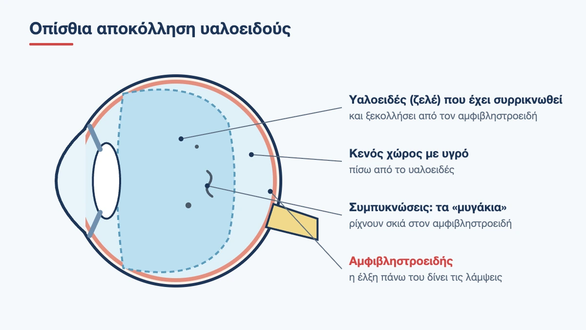
How and why does the vitreous detach?
From our forties onwards the gel starts to change. The hyaluronic acid loses its ability to hold water, the collagen fibres clump together and pockets of liquid form inside the vitreous. In other words, the gel liquefies in the centre and shrinks. At the same time, the “glue” that holds it against the retina weakens.
At some point the liquid finds a way through and passes behind the gel. The vitreous collapses forwards and separates from the retina, first around the macula and last of all from the optic nerve. In its early stages the process can progress silently over months or years, but the final step usually happens suddenly, within hours or days, and that is when the patient notices the symptoms.
The term “posterior vitreous detachment” that you will see on your report means exactly this: the back part of the vitreous has come away. Its front part always remains attached to the periphery of the retina.
Book an appointment with the doctor
An examination and clear advice on your case at our practice in Neo Psychiko.
Vitreous detachment and retinal detachment are not the same
The two names sound alike and this causes a great deal of worry. They are, however, completely different conditions.
| Vitreous detachment | Retinal detachment | |
|---|---|---|
| What comes away | The gel from the retina | The retina itself from the wall of the eye |
| How common | Very common, affects almost everyone with age | Rare, about 1 in 10,000 people a year |
| Symptoms | Floaters, flashes | A shadow or curtain, loss of vision |
| Vision | Not at risk | Seriously at risk |
| Treatment | None, monitoring only | Urgent surgery |
The link between them is that the first can, in a small percentage of cases, cause a tear, and a tear left untreated can lead to the second. You can read more about this serious complication on the page about retinal detachment.
How common is it and who gets it earlier?
Vitreous detachment is rare before the age of 45. In their sixties about 1 in 4 people have it, and after 70 about 2 in 3. It is therefore not a disease but a milestone in the normal ageing of the eye, like presbyopia or grey hair. It appears earlier in people with:
- Myopia: in short-sighted eyes the vitreous liquefies earlier, on average a decade sooner, and the higher the myopia, the earlier it happens
- Cataract surgery: removing the natural lens changes the space and consistency of the vitreous, and many people develop a detachment within the first 1 to 2 years
- Injury to the eye or head
- Intraocular inflammation or bleeding
- Diabetes mellitus
- Menopause: women develop it slightly earlier, probably because the drop in oestrogen affects hyaluronic acid
Symptoms
Floaters
Floaters are shadows cast on the retina by condensations in the vitreous. People describe them as dots, threads, cobwebs or an insect flying in front of the eye. They move with a slight lag when you shift your gaze and are most visible against a bright, even background, such as the sky, a white wall or a computer screen. In acute detachment a large ring-shaped or crescent-shaped floater is typical: it is the spot where the vitreous was attached around the optic nerve, now floating free.
Flashes
Flashes, or photopsia, look like lightning or a camera flash at the edge of your field of vision, usually towards the temple. They are easier to see in the dark and with eye movements. They happen because the vitreous pulls on the retina where it is still attached, and the retina “translates” any mechanical stimulus into light. As long as the flashes continue, there is still traction. Once the vitreous has fully separated, they stop.
Blurred vision
Some patients describe a veil or mist that comes and goes. This is caused by the back surface of the vitreous passing in front of the macula. If the blurring is marked and comes with many small dark specks, there may be a small bleed into the vitreous, which greatly increases the likelihood of a tear.
Posterior vitreous detachment does not cause pain, redness or watering of the eye. In fact, many people go through it without noticing anything at all.
When is it an emergency?
Symptoms alone cannot distinguish a harmless detachment from one that has torn the retina. Only an examination can show that. There are, however, warning signs that make the visit a matter of hours rather than days:
- a sudden “shower” of many small black specks, like soot or a swarm of mosquitoes
- flashes that become more frequent instead of fading
- a shadow, curtain or dark crescent that starts at the edge of your vision and spreads
- a sudden drop in vision
- symptoms in an eye with high myopia, a previous tear, recent surgery or an injury
- a history of retinal detachment in the other eye or in the family
A tear that is found early is treated with laser in the clinic in a few minutes. A tear that is missed can progress to retinal detachment and require surgery. This difference is the reason we never recommend a “wait and see” approach.
Diagnosis
The examination starts with your history: when the symptoms began, whether there are flashes, and whether you have myopia or have had previous eye surgery. Next come measurements of your vision and eye pressure and an examination at the slit lamp. Among other things, we look for pigment granules floating in the anterior vitreous, a finding that strongly suggests a tear is present.
The key step is a dilated fundus examination. The drops take 20 to 30 minutes to work, and then the whole retina is examined, right out to the far periphery, where most tears occur. A special contact lens or gentle pressure on the eyelid is often used to bring the most distant areas into view. Your vision stays blurred for 3 to 4 hours after the drops, so it is best not to drive and to bring sunglasses with you.
Optical coherence tomography (OCT) shows how the vitreous relates to the macula and reveals whether any traction remains at the centre. If bleeding prevents a clear view of the retina, an ultrasound scan of the eye is performed to show whether the retina is in place.
Why is a second examination needed?
A normal first examination is very reassuring, but not final. The vitreous detachment may not yet be complete, and in a small percentage of patients, around 2 to 5%, a tear develops over the following weeks. That is why a follow-up is booked for 4 to 6 weeks later, or sooner if your symptoms change. After this period the risk falls considerably.
Possible complications
- Retinal tear: in about 10 to 15% of acute symptomatic detachments. It is treated with laser around the tear, which “seals” it so that fluid cannot pass underneath
- Vitreous haemorrhage: when a small blood vessel tears. It is usually absorbed, but requires close monitoring
- Retinal detachment: when a tear is left untreated
- Epiretinal membrane: months or years later, from cells that spread over the macula
- Vitreomacular traction and macular hole: when the vitreous stays attached at the centre and pulls on it
In the vast majority of people none of these happen and the detachment completes without any consequences.
Treatment
Vitreous detachment itself cannot be treated and does not need treatment. The gel does not reattach, and there is no reason for it to do so. There are no drops, tablets, vitamins or exercises that affect its course. What we do is make sure the retina is intact and deal immediately with anything we find.
What about the floaters?
For most people the floaters become much less bothersome within 3 to 6 months. There are two reasons for this. The condensations move away from the retina and settle downwards, out of the visual axis, so their shadow becomes fainter. And the brain gradually learns to ignore them, just as it ignores the frame of your glasses or your nose, which is permanently in your field of vision.
A few practical tricks help during this time: sunglasses outdoors, lower brightness or dark mode on screens, and a quick up-and-down eye movement when a floater settles right in the centre, so that it shifts.
When floaters persist
In a small proportion of patients the floaters remain dense and genuinely interfere with reading or driving, even after a year. There are two interventional options, both with advantages and limitations. YAG laser aims to break up large condensations. It is relatively simple, but results vary and it suits only selected cases with a well-defined floater located away from the lens and the retina. Vitrectomy removes the vitreous together with the floaters and is very effective, but it is intraocular surgery with risks, such as cataract and retinal detachment. For this reason it is reserved for a small number of patients with severe, persistent symptoms, after careful discussion.
What to do and what to avoid in the first few weeks
There is no evidence that any activity causes a tear in an eye with vitreous detachment, and bed rest is not needed. As a matter of common sense, while the flashes continue we recommend that you:
- carry on as normal with work, walking, reading and screen use
- avoid, for a few weeks, activities involving strong vibration or sudden jolts of the head, such as martial arts, high diving or heavy weightlifting
- do not rub your eyes hard
- check each eye separately once a day, covering the other, to see whether a shadow has appeared in your peripheral vision
- come in straight away if anything changes, without waiting for your scheduled appointment
Air travel is allowed. The restriction only applies to people who have gas in the eye after surgery.
Book an appointment with the doctor
An examination and clear advice on your case at our practice in Neo Psychiko.
Vitreous detachment after cataract or refractive surgery
People who have had cataract surgery often ask whether floaters appearing months later mean something went wrong. In most cases they do not. The artificial intraocular lens is much thinner than the natural lens, so the vitreous gains more space, moves forwards and detaches earlier than it otherwise would. In addition, once the cloudy lens has been removed, the patient sees more clearly and notices floaters that were already there. A retinal examination is still essential, because the risk of retinal detachment is slightly higher in the first years after surgery, especially in short-sighted eyes and in younger patients.
Laser eye surgery for myopia does not remove the risk that comes with a short-sighted eye. The laser changes the shape of the cornea, not the length of the eye or the condition of the vitreous and retina. Anyone who was short-sighted before surgery needs the same vigilance about floaters and flashes for the rest of their life.
What to mention when you call for an appointment
To judge correctly how soon you need to be seen, it helps to tell us on the phone:
- exactly when the symptoms started and in which eye
- whether you see a few large floaters or many small specks
- whether there are flashes and whether they are becoming more frequent
- whether there is a shadow or an area where you cannot see
- whether you have myopia, recent surgery or a blow to the eye
- whether you or anyone in your family has had a retinal detachment
Come to your appointment with someone or by taxi, as the dilating drops blur your vision for a few hours, and allow about an hour for the visit.
The psychological side: when floaters cause anxiety
Quite a few people, even after learning that their retina is healthy, remain very bothered by their floaters and keep watching them all day. Constantly looking for them makes them more noticeable, because attention works like a magnifying glass. It helps to know that this discomfort is real and common, that it does not signal damage and that, in the vast majority of cases, it eases with time without any intervention. If after several months it still affects your work or your mood, talk about it openly at your next appointment.
Flashes from the eye or from migraine?
Not every flash is a vitreous detachment. The visual aura of migraine is a very different phenomenon: a shimmering zigzag line or a bright arc that slowly expands over 15 to 30 minutes, is seen with both eyes, even when they are closed, and then disappears completely, with or without a headache. Vitreous flashes, by contrast, last a fraction of a second, appear in one eye, in roughly the same position, and recur for days. If you are not sure, a retinal examination settles the question.
Will it happen in the other eye?
In all likelihood, yes. Because both eyes are the same age and have the same make-up, the second eye usually follows within 1 to 3 years. If there was a tear in the first eye, the chance of the same happening in the second is higher, so being alert to symptoms and getting examined promptly matter even more. The good news is that the second time you already know what the symptoms mean and how quickly you need to act.
When to book an appointment
If you have seen floaters or flashes for the first time, or if your old floaters have suddenly multiplied, you need a dilated retinal examination within 1 to 2 days. If there is a shadow or a drop in vision, the same day. At the clinic in Neo Psychiko, Athens, we see these cases as a priority. Call 210 677 0007 and describe your symptoms so that we can see you promptly. Read also the article on floaters and retinal tears.
Suddenly seeing floaters or flashes?
A dilated retinal examination within 24 to 48 hours shows whether there is a tear. Call for an urgent appointment.
Frequently Asked Questions about Posterior Vitreous Detachment
Is posterior vitreous detachment dangerous?
Not in itself. It is a normal age-related change and does not harm your sight. What can be dangerous is its complication, a retinal tear, which occurs in about 1 in 10 patients with sudden symptoms. That is why a dilated retinal examination is needed within 24 to 48 hours.
How long do the flashes and floaters last?
The flashes usually settle within a few weeks, once the vitreous has fully separated. The floaters become noticeably less bothersome within 3 to 6 months, as they settle and the brain learns to ignore them. They rarely disappear completely.
Can posterior vitreous detachment be cured?
There is no treatment and none is needed, because the vitreous does not have to stay attached. No eye drop, vitamin or exercise changes its course. Only the complications need treatment, such as a retinal tear, which is treated with laser.
Why do I need a follow-up if my first examination was clear?
Because the detachment may not have been complete on the day of the first examination. In 2 to 5% of patients a tear appears over the following weeks. A follow-up at 4 to 6 weeks, or sooner if your symptoms change, covers this period of risk.
Can I exercise and travel by plane?
Flying is allowed without restriction. So are everyday activities and walking. For a few weeks, while the flashes continue, it is sensible to avoid contact sports and exercise involving sharp jolts of the head.
Is it caused by stress, tiredness or screens?
No. The cause is the natural liquefaction of the vitreous with age, which is accelerated by myopia, cataract surgery or an injury. Stress and tiredness may make you notice the floaters more, but they do not create them.
If I have had floaters for years, should I be worried?
Old, stable floaters are usually harmless. What warrants concern is a change: a sudden increase in their number, new flashes or a shadow in your peripheral vision. In that case you need an urgent examination, even if you have been checked before.
Medically reviewed by: Dr. Vasileios Skouteris MD, FEBO