
Written by Vasileios Skouteris MD, FEBO, Ophthalmic Surgeon
Page contents
- What is xanthelasma?
- How common is it, and who gets it?
- Causes and risk factors
- Xanthelasma and the heart: why it is not just a cosmetic issue
- What can it be confused with?
- Diagnosis and check-up
- Do diet or cholesterol medicines make them go away?
- Xanthelasma removal methods
- Preparing for removal
- Xanthelasma in children and young adults
- Until removal: covering with make-up
- How the ophthalmologist and the cardiologist work together
- After removal: recovery and care
- Risks and recurrence
- What not to do yourself
- Prevention and long-term care
- When to book an appointment
- Frequently Asked Questions about Xanthelasma
Xanthelasma are soft, yellowish plaques that appear on the skin of the eyelids, usually at the inner corner near the nose. They are made of cholesterol deposits within the skin, are benign and do not affect vision. They matter for two reasons, though: in roughly half of people they are linked with a lipid disorder and a higher cardiovascular risk, and they never go away on their own. Removal is done for cosmetic reasons, using laser, surgical excision or a chemical peel.
What is xanthelasma?
Xanthelasma is the most common form of xanthoma, a fatty deposit in the skin. The name comes from the Greek words “xanthos” (yellow) and “elasma” (plate), a yellow plaque, which describes exactly what we see. Under the microscope, the upper layer of the dermis contains clusters of immune cells that have “swallowed” cholesterol and filled up with fat droplets, which is why they are called foam cells. This is the same type of cell found in atherosclerotic plaques in the arteries, and that similarity explains why xanthelasma is also of interest to cardiologists.
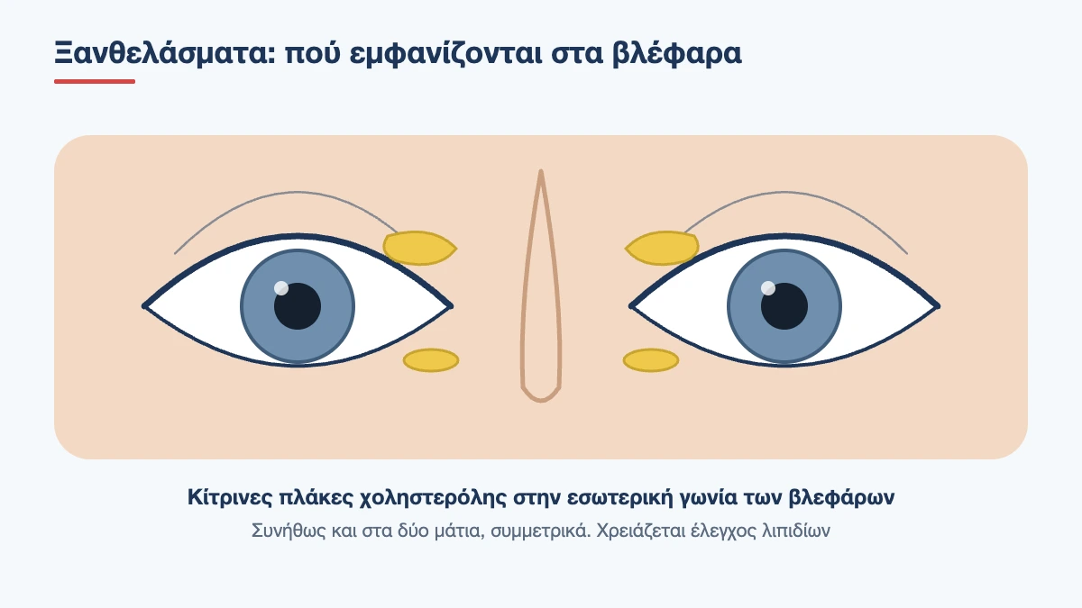
Why they appear specifically on the eyelids is not entirely clear. The skin there is the thinnest in the body, moves thousands of times a day as we blink and has a rich network of tiny blood vessels, from which lipids seem to leak more easily into the tissues.
What they look like
- yellow, yellowish-white or orange in colour
- flat or slightly raised, with well-defined edges
- soft or semi-firm to the touch, as if there is something under the skin
- they usually start at the inner corner of the upper eyelid. The lower eyelid follows
- as a rule they appear on both eyes, almost symmetrically
- size ranges from 2 millimetres to plaques covering a large part of the eyelid
- they grow slowly, over months and years, and neighbouring plaques may merge
They do not hurt, itch or become inflamed, and they never turn malignant. Only when they become very large can they make the eyelid feel heavy. The problem they cause is cosmetic, and for many people this matters, because they sit on the most visible part of the face and are not easy to cover with make-up.
How common is it, and who gets it?
It is estimated that 1 to 4 in every 100 adults have xanthelasma. It usually appears between the ages of 40 and 60, slightly more often in women. When it appears before the age of 40, and especially in children or teenagers, it often points to an inherited lipid disorder and must always be investigated.
Book an appointment with the doctor
An examination and clear advice on your case at our practice in Neo Psychiko.
Causes and risk factors
Lipid disorders
About half of people with xanthelasma have raised cholesterol or triglycerides in the blood. The most common abnormalities are:
- high “bad” cholesterol (LDL)
- low “good” cholesterol (HDL)
- raised triglycerides
- familial hypercholesterolaemia, an inherited condition in which LDL is very high from childhood and a heart attack can occur early if treatment is not started
Conditions that raise lipids secondarily
- hypothyroidism
- diabetes mellitus
- chronic liver diseases with bile stasis, such as primary biliary cholangitis
- nephrotic syndrome
- obesity and metabolic syndrome
- certain medicines, such as corticosteroids, oestrogens, oral retinoids and some antiretrovirals
When blood lipids are normal
The other half have normal test results. In these people, inherited predisposition, local factors in the eyelid skin and subtler abnormalities in the way the body transports and clears cholesterol, which do not show up in routine tests, seem to play a part. Smoking, high blood pressure and a family history of xanthelasma increase the likelihood.
Xanthelasma and the heart: why it is not just a cosmetic issue
This is the most important message on this page. Large population studies, which followed tens of thousands of people for decades, have shown that people with xanthelasma have heart attacks and coronary artery disease more often than those without. The finding held even when blood cholesterol was normal, and regardless of smoking, blood pressure and weight.
This does not mean that anyone with xanthelasma will have a heart attack. It means that the skin is giving a clue about how the body handles cholesterol, and that a full check of cardiovascular risk factors is worthwhile. In this sense xanthelasma is useful: it is a visible, painless sign that can lead to early prevention.
A related finding, which we see during the slit lamp examination, is arcus senilis, a whitish ring at the edge of the cornea caused by lipid deposits. In older people it is normal. Under the age of 50, especially alongside xanthelasma, it strengthens the suspicion of hyperlipidaemia.
What can it be confused with?
The diagnosis is usually made at first glance. There are, however, other eyelid lesions with a yellowish or whitish appearance:
- Syringomas: many small, firm, skin-coloured bumps, mainly on the lower eyelids. They are benign tumours of the sweat glands
- Milia: tiny, white, hard keratin cysts that look like grains
- Sebaceous hyperplasia: yellowish bumps with a small dip in the centre
- Chalazion: a firm lump within the thickness of the eyelid rather than on the skin surface, usually single
- Rare xanthogranulomas: firmer, more extensive and deeper plaques, sometimes ulcerated, which are associated with blood disorders. A firm texture, rapid growth or spread beyond the eyelids are reasons for a biopsy
Diagnosis and check-up
The diagnosis is clinical and a biopsy is rarely needed. What matters is the check-up that follows. For everyone with xanthelasma we recommend, in cooperation with a GP, internist or cardiologist:
- A full lipid profile: total cholesterol, LDL, HDL and triglycerides, after fasting
- Blood glucose and glycated haemoglobin (HbA1c)
- Thyroid function (TSH)
- Liver enzymes and markers of cholestasis
- Kidney function tests
- Blood pressure measurement and assessment of overall cardiovascular risk
In younger patients, or when there is a family history of early heart disease, the check-up is extended to more specialised tests, such as lipoprotein(a), and testing of first-degree relatives is recommended.
During the eye examination we record the size, thickness and position of each plaque, how much excess skin there is on the eyelid and how close the lesions are to the tear duct opening (punctum) and the eyelid margin. These factors determine which removal method is safe.
Do diet or cholesterol medicines make them go away?
Usually not. Controlling lipids with diet, exercise and medication is essential for the heart and blood vessels, and appears to slow the growth of the plaques and reduce the chance of them coming back after removal. Plaques that have already formed, however, rarely shrink noticeably. There are isolated reports of regression after many months of treatment, but this is not something you can count on. If you want them gone, local treatment is needed.
Xanthelasma removal methods
There is no single best method for everyone. The choice depends on the size and depth of the plaque, its position, skin colour, whether there is excess eyelid skin and the doctor’s experience.
| Method | Best suited to | Advantages | Disadvantages |
|---|---|---|---|
| Surgical excision | Large, thick or deep plaques, especially when there is excess skin | Removes the full thickness, provides tissue for examination, can be combined with blepharoplasty | Stitches, a fine scar, risk of pulling the eyelid if too much skin is removed |
| Laser | Small to medium, superficial plaques | Precise, bloodless, no stitches, quick healing | Possible change in skin colour, sometimes two sessions are needed |
| Chemical peel with trichloroacetic acid | Small, flat plaques | Simple, quick, low cost | Risk of loss of skin colour or scarring, more frequent recurrence |
| Radiofrequency | Small plaques close to the eyelid margin | Good control of depth | Highly dependent on the operator |
| Cryotherapy | Rarely | Simple | Marked eyelid swelling, frequent loss of skin colour |
Laser removal
The laser vaporises the plaque layer by layer until healthy dermis is reached, while sealing the tiny blood vessels at the same time, so there is no bleeding. It is done under local anaesthetic, with a cream or a small injection, and takes 10 to 20 minutes. The eyes are protected with special metal shields. A scab forms over the area and falls off in 7 to 10 days, leaving pink skin that returns to its normal colour within a few weeks. It is the same technology used in laser blepharoplasty.
Surgical excision
The plaque is removed in its full thickness with a scalpel or a cutting laser, and the skin is closed with very fine stitches, which are removed after 5 to 7 days. When the eyelid has loose skin, the excision is planned as part of a blepharoplasty: in one procedure both the xanthelasma and the excess skin are removed, and the scar is hidden in the natural crease. The lower eyelid needs more care, because removing skin there can pull the eyelid downwards. For very extensive lesions, removal is done in stages or with a small skin graft.
Chemical peel
A high-concentration trichloroacetic acid solution is applied with a fine applicator directly onto the plaque, which turns white immediately. A scab follows and healing takes about two weeks. Repeat treatments are often needed. This is a medical procedure and not something that can be done at home.
Preparing for removal
Removal is a planned procedure, so there is time to do it properly. Before the day of the procedure:
- complete your blood tests and, if needed, start treatment to control your lipids. The better controlled they are, the lower the chance of recurrence
- tell us about all your medicines, especially anticoagulants, aspirin and oral retinoids. Do not stop anything without instructions
- mention whether you tend to develop hypertrophic scars or keloids, or whether your skin darkens easily after an injury
- avoid intense sunbathing for the two weeks beforehand. Tanned skin has a higher risk of colour change
- choose autumn or winter if you can, when sun protection is easier
- come without make-up and without contact lenses
- if there is a social event you want to look your best for, schedule the removal at least three weeks earlier
A photograph of your eyelids before the procedure is useful, both for comparing the result and for spotting a small recurrence early, which is easier to treat while it is still small.
Xanthelasma in children and young adults
Xanthelasma is rare in children and, when it does occur, should never be regarded as a simple cosmetic issue. It is often the first visible sign of familial hypercholesterolaemia, an inherited condition affecting about 1 in 250 people, in which early treatment radically changes the outlook. Alongside xanthelasma there may be deposits in the tendons, mainly the Achilles tendon and the finger joints. The child is referred to a paediatrician or a lipid specialist, and parents and siblings are tested. The same applies to adults under 40, especially if there is a family history of heart attack or stroke at a young age.
Book an appointment with the doctor
An examination and clear advice on your case at our practice in Neo Psychiko.
Until removal: covering with make-up
For those who do not want, or are not yet able, to go ahead with removal, flat plaques can be covered. You need a colour corrector in a peach or lilac shade, which neutralises the yellow, followed by a concealer that matches your skin tone, applied with light dabs rather than rubbing. Choose ophthalmologically tested products and remove them gently in the evening, without pulling the eyelid. Raised plaques are harder to cover, because the relief shows up in side lighting.
How the ophthalmologist and the cardiologist work together
Care has two parts that run in parallel. The ophthalmologist makes the diagnosis, checks the eyes for other signs of lipid deposits and carries out the removal, if you want it. The internist or cardiologist assesses your overall risk, taking into account age, blood pressure, smoking, blood sugar and family history, and decides whether lifestyle changes are enough or whether medication is needed. Some people are also asked to have imaging of their blood vessels. In other words, xanthelasma is the starting point for an overall assessment that goes far beyond the eyelids.
After removal: recovery and care
- cold compresses for the first 24 hours to reduce swelling
- antibiotic ointment on the area for one week
- do not scratch or pick off the scab, it will fall off on its own
- no make-up on the area until the skin has fully healed
- high-protection sunscreen and sunglasses for at least 2 to 3 months, because new skin darkens easily in the sun
- no swimming or sauna for two weeks
- back to work the next day, if the appearance of the scab does not bother you
Risks and recurrence
Whatever the method, the possible side effects are similar: a mark lighter or darker than the surrounding skin, scarring, prolonged redness and, rarely, pulling of the eyelid if healing involves marked contraction. Colour changes are more common in darker skin and usually improve with time.
The issue you need to know about before deciding is recurrence. Xanthelasma comes back in a significant proportion of cases, reaching up to 40% after the first removal in studies, usually within the first year. The chance is higher when:
- blood lipids remain uncontrolled
- there are plaques on all four eyelids
- the lesion was deep and was not removed in its full thickness
- there has already been one recurrence
- there is inherited hypercholesterolaemia
Recurrence is not a failure of the procedure. The body’s tendency to deposit cholesterol in this area remains. If it happens, removal can be repeated.
What not to do yourself
The internet is full of “natural” remedies using garlic, vinegar, castor oil, lemon juice or acid creams. None has been shown to dissolve xanthelasma, and several have caused chemical burns, permanent scars and loss of skin colour on skin half a millimetre thick, a few centimetres from the cornea. Do not try to prick or squeeze them either. They are not cysts with contents that can be emptied: the fat sits inside the skin cells.
Prevention and long-term care
- bring your cholesterol and triglycerides to the targets set by your doctor, and do not stop treatment once your values improve
- follow a Mediterranean-style diet, low in saturated fat and processed foods
- exercise regularly and keep your weight at a healthy level
- stop smoking
- keep diabetes, thyroid function and blood pressure under control
- repeat your lipid tests once a year, even if the first result was normal
When to book an appointment
If you have noticed yellow plaques on your eyelids, an examination confirms the diagnosis, rules out other lesions and points you to the check-up you need. If you are considering removal, we discuss which method suits the size and position of the lesions and what result is realistic. See also the other cosmetic treatments for the eye area. Call the clinic on 210 677 0007 or use the contact form.
Yellow plaques on your eyelids?
We examine the lesions and recommend the right removal method for you. Book an appointment for an assessment.
Frequently Asked Questions about Xanthelasma
Is xanthelasma dangerous?
Not in itself. Xanthelasma plaques are benign cholesterol deposits in the skin and do not affect the eyes or vision. However, they are often linked with raised blood lipids and a higher cardiovascular risk, so blood tests and an assessment by a GP, internist or cardiologist are needed.
Will they go away on their own if I lower my cholesterol?
As a rule, no. Controlling blood lipids protects the heart, slows the growth of the plaques and reduces recurrence, but existing plaques rarely shrink. Removing them requires local treatment.
What is the best removal method?
It depends on the lesion. Small, superficial plaques respond well to laser. Large, thick or deep plaques are better treated with surgical excision, which is often combined with blepharoplasty. The choice is made after the examination.
Is removal painful, and does it leave a mark?
It is done under local anaesthetic and is not painful. A scab forms over the area for 7 to 10 days. A fine mark or a slight difference in skin colour may remain, which usually improves within a few months with sun protection.
Do they come back after removal?
They can. Recurrence rates reach up to 40%, usually within the first year. Recurrence is more common when blood lipids remain uncontrolled, when all four eyelids are affected or when the plaque was deep. Removal can be repeated.
I have xanthelasma but my cholesterol is normal. What does that mean?
This is the case in about half of patients. Even with normal values, studies show a slightly higher cardiovascular risk, so a more thorough check-up and repeating the blood tests every year are worthwhile.
Can I remove them at home with garlic or acids?
No. No home remedy has been shown to work, and burns, scarring and permanent loss of skin colour have been reported. Eyelid skin is the thinnest in the body and sits right next to the eye, so removal must be carried out by a doctor.
Medically reviewed by: Dr. Vasileios Skouteris MD, FEBO