Page contents
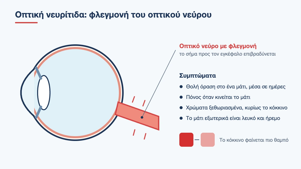
Optic neuritis is inflammation of the optic nerve, the “cable” that carries the image from the eye to the brain. It usually affects adults aged 20 to 45, more often women, and shows up as blurred vision in one eye that gets worse over a few days, pain when the eye moves and colours that look washed out. In most cases vision recovers to a large extent within weeks. It does, however, need prompt investigation, because it can be the first sign of a neurological condition.
What Is the Optic Nerve and What Happens to It?
The optic nerve is made up of about one million nerve fibres that start in the retina and end in the brain. Each fibre is surrounded by an insulating sheath, myelin, which allows the electrical signal to travel quickly, like the insulation on a cable. In typical optic neuritis, the immune system mistakenly attacks this myelin. The nerve swells, the insulation is damaged in patches and the signal slows down or is interrupted. The image reaches the brain late, weak and distorted.
Myelin can regenerate to a significant degree, and this is why vision usually recovers. The repair is not perfect, however, which is why many patients notice that, although they can read the chart again, the eye is not quite the same as before.
Symptoms of Optic Neuritis
- Blurred vision in one eye, which gets worse over hours to days and reaches its lowest point in 1 to 2 weeks. Severity ranges from slight dimness to almost complete loss
- Pain behind or around the eye, which gets worse when you move it. It is present in 9 out of 10 patients and often comes a few days before the blurring
- Washed-out colours. Red looks pink or orange, and everything looks more “faded” compared with the other eye
- A dark or blurred patch in the centre of the field of vision
- Reduced contrast and brightness. The eye sees as if the lights have been dimmed
- Flashes or sparks with eye movements, in some people
- Worsening with heat. After a hot bath, exercise or a fever, vision blurs temporarily, and it returns once body temperature falls
From the outside the eye looks completely normal: white, with no discharge and no swelling. This sets it apart from other causes of eye pain, where there is usually redness.
A simple test at home
Look at a bright red object, for example a bottle cap, first with one eye and then with the other. If it looks clearly duller or paler with one eye, the optic nerve of that eye is probably not working properly. The test does not replace an examination, but it is a good reason not to put one off.
Causes of Optic Neuritis
Demyelinating optic neuritis
This is the most common form in young adults, the one described above. It may occur on its own, never to be followed by anything else, or it may be the first sign of multiple sclerosis, a condition in which the same immune attack on myelin also happens in other parts of the brain and spinal cord. In about 1 in 5 patients with multiple sclerosis, optic neuritis was the first symptom.
Other autoimmune forms
Two rarer conditions, detected by specific antibodies in the blood, cause optic neuritis that behaves differently: more severe loss of vision, often in both eyes, less spontaneous recovery and a tendency to relapse. They need more aggressive and long-term treatment, which is why recognising them early is so important.
Infections and systemic diseases
Optic neuritis can accompany viral infections, especially in children, where it often affects both eyes and has a good outlook. More rarely it is caused by syphilis, Lyme disease, tuberculosis, sarcoidosis or lupus.
What is not optic neuritis
Some optic nerve conditions look similar but are not inflammation and are treated differently:
- Ischaemic optic neuropathy: a “stroke” of the optic nerve. It affects people over 50, the loss is sudden, usually noticed on waking in the morning, and it is painless. When there is also a headache at the temples and pain when chewing, giant cell arteritis must be ruled out immediately
- Toxic and nutritional neuropathy: slow, painless loss in both eyes, from certain medicines, alcohol and tobacco misuse or vitamin B12 deficiency
- Compression of the nerve by a tumour or by enlarged muscles in thyroid eye disease: slow, progressive worsening
- Hereditary neuropathies: painless loss in young men, first in one eye and then in the other
- Glaucoma: chronic damage to the nerve from eye pressure, without symptoms for years
Diagnosis
Eye examination
- Visual acuity and colour vision with special charts
- Pupil testing. When we shine a light alternately into each eye, the pupil of the affected eye dilates instead of constricting. This is the most objective sign of optic nerve damage and is present even when vision is relatively good
- Fundoscopy. In two thirds of patients the nerve looks normal, because the inflammation lies behind the eye. In the rest, the head of the nerve is swollen
- Visual field testing, which maps the area where vision has been lost and is used to monitor recovery
- Optical coherence tomography (OCT), which measures the thickness of the nerve fibres, rules out macular conditions with similar symptoms and shows months later whether any thinning remains
MRI scan
An MRI scan of the brain and orbits with contrast is the most important test after diagnosis. It confirms inflammation of the nerve and, above all, shows whether there are areas of demyelination in the brain. This finding determines the outlook and the follow-up that comes next.
Blood tests
Tests include markers of inflammation, screening for infections and autoimmune diseases, and the specific antibodies for the atypical forms, especially when the loss of vision is severe, affects both eyes or does not improve as expected. In some cases the neurologist completes the investigation with a lumbar puncture.
Treatment of Optic Neuritis
In the typical form, vision starts to improve on its own within 2 to 4 weeks, whether or not treatment is given. The large clinical trial that shaped how it is managed showed three things:
- high-dose intravenous steroids for three days speed up recovery by a few weeks
- they do not change final vision at six months or one year
- oral steroids at a standard dose, without intravenous steroids first, were linked to more relapses and should not be given on their own
The decision to treat is therefore made case by case. Intravenous steroids are preferred when the loss of vision is severe, when both eyes are affected, when the other eye already sees poorly, when the patient needs a quick recovery for work reasons or when the MRI shows lesions. In the atypical autoimmune forms, treatment is essential and immediate, often with plasma exchange and long-term immunosuppression.
If the investigation shows an increased risk of multiple sclerosis, the neurologist may suggest starting disease-modifying treatment, which reduces the likelihood and frequency of new episodes.
How Long It Lasts and What to Expect
- First 1 to 2 weeks: vision gets worse and pain is at its peak
- Weeks 2 to 5: the pain settles and improvement begins
- Months 1 to 3: most of the recovery
- Up to 12 months: slow, further improvement
More than 9 out of 10 patients with typical optic neuritis regain very good visual acuity. Subtle differences often remain, however: colours slightly duller, reduced contrast in low light, difficulty judging movement and temporary blurring with heat or tiredness. These differences do not mean new inflammation.
If vision has not started to improve after 4 to 5 weeks, or if it keeps getting worse after the first two, the diagnosis should be reviewed.
Optic Neuritis and Multiple Sclerosis
This is the question on every patient’s mind, and it deserves an honest answer. Optic neuritis does not mean you have multiple sclerosis, but it does increase the chance of it developing in the future. By how much depends mainly on the MRI scan at the time of the first episode. In long-term follow-up of patients over 15 years:
- when the brain MRI was normal, about 1 in 4 developed the disease
- when there was even one lesion, about 7 in 10
These figures come from a time before modern treatments. Today multiple sclerosis is diagnosed earlier and treated with medicines that substantially change its course. In this sense, optic neuritis acts as an early warning that allows monitoring to begin at the right time.
What You Can Do
- do not drive while the vision in one eye is significantly reduced, because depth perception is affected
- avoid overheating, such as saunas, very hot baths and strenuous exercise in the heat, if you notice that your vision blurs. This blurring is temporary and does not harm the nerve
- stop smoking, which is linked to a worse course
- have your vitamin D levels checked with your doctor
- keep your appointments with the neurologist, even if your vision has fully recovered
- report any new symptom straight away: blurring in the other eye, double vision, numbness, weakness, unsteadiness or bladder problems
When to Be Examined Urgently
Any blurring in one eye that gets worse over days, especially together with pain on eye movement or a change in colour vision, needs an examination within 24 hours. If the loss is sudden and painless, if you are over 50 or if you have a headache and jaw pain, it needs to be seen the same day. At the clinic in Neo Psychiko, Athens, a full eye examination with visual fields and OCT is carried out, and collaboration with a neurologist for the MRI and treatment is arranged without delay. Call 210 677 0007. See also the article on the causes of blurred vision.
Frequently Asked Questions about Optic Neuritis
Will my vision come back after optic neuritis?
In the typical form, more than 9 out of 10 patients regain very good vision. Improvement starts within 2 to 4 weeks and continues for up to a year. A small difference in colours and contrast often remains.
Does optic neuritis mean I have multiple sclerosis?
Not necessarily. Many people have a single episode and nothing else. It does increase the risk, however, and by how much depends on the brain MRI: the risk is lower when it is normal and higher when there are lesions.
Are steroids always needed?
No. Intravenous steroids speed up recovery but do not change final vision in the typical form. They are recommended when the loss is severe or affects both eyes. In the atypical autoimmune forms they are essential and urgent.
How long does an episode last?
Vision worsens for 1 to 2 weeks, stays stable for a short while and then improves. Most of the recovery happens in the first 3 months. The pain usually settles within a week.
Why does my vision blur when I get hot?
When body temperature rises, nerve fibres that have lost some of their myelin transmit the signal less easily. The blurring is temporary, settles once you cool down and does not mean new damage.
Can it happen again?
Yes, in the same or the other eye, in about 1 in 3 patients within ten years. Repeated relapses need testing for the atypical autoimmune forms and a discussion with a neurologist about preventive treatment.
Is it caused by stress or screens?
No. It is an autoimmune inflammation of the nerve and is not related to eye strain, reading or screens. Stress does not cause it, although a difficult period may coincide with its onset.
Questions about your case?
Dr. Skouteris will examine you at his practice in Neo Psychiko and explain your options.

Ophthalmic surgeon trained at the John Radcliffe University Hospital in Oxford and Fellow of the European Board of Ophthalmology (FEBO).
Full profileRelated articles
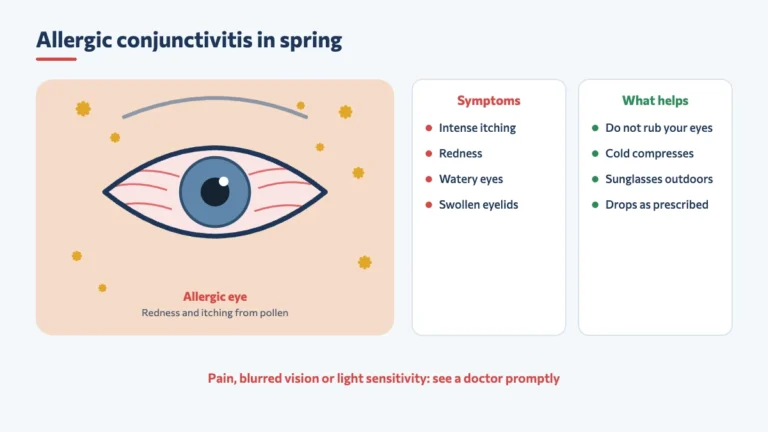
Allergic Conjunctivitis in Spring: Symptoms, Prevention and When to Worry
Itching, redness and watering in both eyes every spring? How to recognise allergic conjunctivitis, what helps prevent it and which signs need prompt examination.
Read the article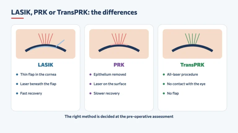
LASIK, PRK or TransPRK: Which Laser Eye Surgery Is Right for You?
The difference between LASIK, PRK and TransPRK, how long recovery takes and which criteria determine the right laser eye surgery for each eye.
Read the article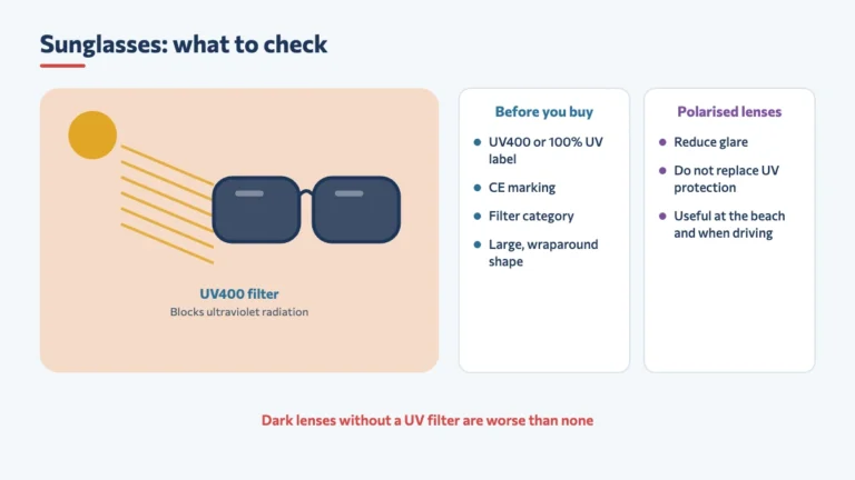
Sunglasses and Eye Protection: What to Check Before You Buy (Including for Children)
UV400, CE mark, filter category and polarised lenses: what to check before buying sunglasses for yourself and for your children.
Read the article