
Written by Vasileios Skouteris MD, FEBO, Ophthalmic Surgeon
Page contents
- How do tears normally drain?
- A watery eye: is the tear duct always to blame?
- Congenital blocked tear duct in babies
- Acquired blocked tear duct in adults
- Symptoms in adults
- Diagnosis
- Treatment in adults
- After dacryocystorhinostomy
- What you can do until the definitive treatment
- When to get examined
- Frequently Asked Questions about Blocked Tear Ducts
A blocked tear duct is a partial or complete blockage of the system that drains tears from the eye into the nose. The tears have nowhere to go, so they pool and spill over onto the cheek, and the eye often produces mucus and suffers repeated infections. It is very common in babies, where it usually opens on its own within the first year, and in adults over 50, where the definitive solution is, as a rule, surgical.
How do tears normally drain?
Tears are produced continuously by the lacrimal gland, which sits under the outer part of the upper eyelid, and by many small glands in the eyelids and conjunctiva. They spread across the surface of the eye with every blink; some of them evaporate and the rest must drain away. The drainage route has four parts:
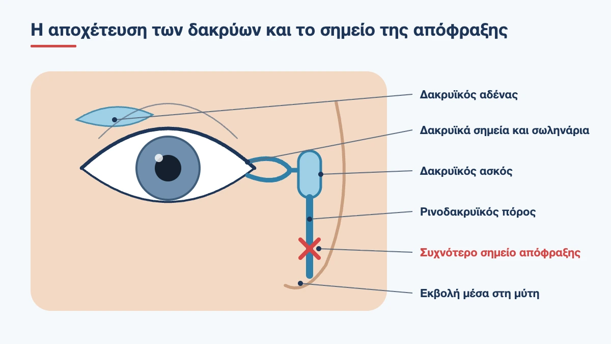
- The puncta: two tiny openings, one on the upper and one on the lower eyelid, at the corner nearest the nose. You can see them with the naked eye if you gently pull down the lower eyelid
- The canaliculi: two thin channels about one centimetre long that start at the puncta and join together
- The lacrimal sac: a small reservoir inside a bony hollow at the side of the bridge of the nose
- The nasolacrimal duct: the tube that runs down through the bone and opens inside the nose, under the inferior turbinate
This is why your nose runs when you cry, and why you sometimes taste eye drops in your throat. Drainage does not happen through gravity alone. Every blink works like a pump: the eyelid muscle squeezes the canaliculi and the sac and pushes the tears downwards. This means the eye can water either because the tube is blocked or because the pump is not working, for example when the lower eyelid is lax.
A watery eye: is the tear duct always to blame?
No, and this is the first thing to clarify, because the treatment is completely different. There are two broad categories:
- Too many tears are produced. The eye becomes irritated and waters reflexively. The most common cause, surprisingly, is dry eye: the surface dries out, the eye “orders” emergency tears and they overflow. Blepharitis, allergy, an ingrowing eyelash or a foreign body do the same
- Tears do not drain. Production is normal, but somewhere along the route there is a narrowing or blockage, or the eyelid does not sit properly against the eye
A few clues point the way. When drainage is the problem, tears run down the cheek even indoors, the eye is often stuck together in the morning and the problem usually affects one eye. When irritation is the problem, there is burning or a gritty feeling, the watering gets worse when reading, using screens and in the wind, and it usually affects both eyes. The two conditions often occur together. For a broader overview, see the article why does my eye water.
Book an appointment with the doctor
An examination and clear advice on your case at our practice in Neo Psychiko.
Congenital blocked tear duct in babies
Why it happens
The nasolacrimal duct forms in the womb as a solid cord that gradually hollows out from top to bottom. The last point to open is where it enters the nose, and in many newborns a thin membrane remains there. It is estimated that about one in ten babies, and according to some studies up to one in five, has some degree of blockage in the first weeks. It is not a malformation, nor is it caused by anything the mother did wrong during pregnancy.
What it looks like
Symptoms usually start in the second to third week of life, when normal tear production begins:
- the eye is constantly wet, with tears running down without the baby crying
- yellowish sticky discharge, especially after sleep, and lashes that stick together
- the white of the eye stays white. This is what distinguishes it from conjunctivitis
- irritated skin on the eyelids from the constant moisture
- symptoms get worse with cold, wind and a runny nose
What we do in the first year
In nine out of ten children the membrane opens on its own by their first birthday, most of them within the first six months. That is why the initial approach is conservative:
- Cleaning: wipe away the discharge with gauze soaked in cooled boiled water or saline, from the nose outwards
- Tear sac massage: with clean hands and short nails, place your index finger on the side of the nose, right next to the corner of the eye, press firmly and slide downwards along the nose. Five to ten strokes, three to four times a day, for example at every nappy change. The pressure created inside the duct helps to break the membrane. Ask us to show you the movement in the clinic, because simply stroking the skin has no effect
- Antibiotic drops: only when the discharge becomes thick and pus-like, for a few days. They do not open the duct and there is no point in using them continuously
When probing is needed
If symptoms persist at around 12 months, probing is scheduled. A very fine metal probe is passed from the punctum down to the nose and opens the membrane. It is done under brief general anaesthesia, takes a few minutes and the child goes home the same day. Success exceeds 90% when it is done in good time and falls as the child gets older, so we do not recommend waiting beyond 12 to 15 months. If the first probing does not work, it can be repeated with balloon dilation of the duct or with placement of a thin silicone tube for a few months.
When a baby’s watery eye is urgent
- red, hot, firm swelling between the eye and the nose, with or without fever: this is an infection of the tear sac
- a bluish lump in the same place from birth: a cyst of the sac, which can also make breathing difficult
- watering together with marked sensitivity to light and a cornea that looks large or cloudy: congenital glaucoma, a rare but serious condition, must be ruled out
- a red eye with a lot of pus in the first days of life
Acquired blocked tear duct in adults
In adults the blockage develops slowly, over months or years, and rarely clears on its own. The cause depends on where along the route the problem lies.
Punctal stenosis
The small openings narrow or close with age, chronic blepharitis, long-term use of certain eye drops, for example for glaucoma, or after viral infections. Often the punctum is open but has moved away from the eye because the lower eyelid has loosened and turned outwards (ectropion), so it cannot “drink” the tears.
Canalicular blockage
This is caused by injury to the corner of the eye, herpes infections, certain chemotherapy drugs or chronic infection of the canaliculus itself. The latter produces a very characteristic red, swollen punctum with yellowish discharge and is often mistaken for months for conjunctivitis that will not clear up.
Nasolacrimal duct obstruction
This is the most common form. The duct gradually narrows from chronic mild inflammation and fibrosis, with no clear cause. It occurs mainly after the age of 50 and is clearly more common in women, probably because their bony canal is anatomically narrower. Other causes include chronic rhinitis and sinusitis, nasal polyps, fractures of the nose and face, previous nasal surgery, radioactive iodine treatment, radiotherapy and, rarely, a tumour. Blood in the tears or a hard lump above the level of the corner of the eye always requires imaging.
Symptoms in adults
- constant watering that runs down the cheek and gets worse in cold and wind
- blurred vision from the pool of tears, especially when reading and driving
- mucous or pus-like discharge and eyelids that stick together in the morning
- “conjunctivitis” that comes back as soon as the drops are stopped
- irritation and eczema on the skin of the lower eyelid from constant wiping
- a soft swelling at the corner near the nose that releases mucus from the punctum when pressed
- fogging of glasses and social discomfort, because people think you are crying
Acute dacryocystitis
When the sac is blocked, its stagnant contents easily become infected. Acute infection shows up as severe pain, redness and firm swelling between the eye and the nose, which can spread to the cheek and come with a fever. It needs oral antibiotics started immediately, and sometimes drainage of the abscess. The infection settles, but as long as the duct remains closed it will keep coming back, so the definitive operation is planned after the episode.
A blocked and infected sac is also a source of bacteria next to the eye. That is why, before any intraocular surgery such as cataract surgery, a blockage with discharge must be dealt with.
Diagnosis
The examination takes place in the clinic and needs no preparation.
- Slit lamp examination: we check the height of the tear lake, the position and opening of the puncta, laxity of the lower eyelid, tear quality and the presence of blepharitis or another cause of irritation
- Fluorescein dye disappearance test: a drop of fluorescein is placed in both eyes. Normally it has almost gone within five minutes. If it remains strong in one eye, drainage on that side is lagging. This is the key test in children too, because it causes no discomfort at all
- Lacrimal syringing: after an anaesthetic drop, a fine blunt cannula is inserted into the punctum and saline is flushed through. If you feel it in your throat, the route is open. If it comes back, there is a blockage, and where it comes back from tells us at what level it lies. The test is slightly uncomfortable but not painful
- Nasal examination: rhinoscopy or endoscopy, often in collaboration with an ENT specialist, for a deviated septum, polyps or inflammation
- Imaging: a CT scan or a special contrast X-ray (dacryocystogram), when there is a history of trauma, a suspected tumour or a previous failed operation
Book an appointment with the doctor
An examination and clear advice on your case at our practice in Neo Psychiko.
Treatment in adults
Drops, massage and flushing do not open a duct that has closed from fibrosis. Antibiotics reduce the discharge temporarily but do not solve the problem. Treatment depends on the site of the blockage.
Punctoplasty (opening the puncta)
When the problem is only a narrow opening, it is widened with a minor procedure lasting a few minutes under local anaesthesia in the clinic. If the lower eyelid is lax or turned outwards, it is corrected by tightening, so that the punctum returns to its proper position and the pump can work again.
Dacryocystorhinostomy (DCR)
When the nasolacrimal duct is blocked, the solution is to bypass it. In dacryocystorhinostomy a new, direct opening is created between the tear sac and the inside of the nose by removing a small piece of the bone that separates them. There are two approaches:
- External: through an incision of about one centimetre on the side of the nose, which heals almost invisibly. This is the classic method, with success rates above 90%
- Endoscopic: entirely through the nose, with an endoscope, with no skin incision at all. It offers faster recovery, allows nasal problems to be corrected at the same time and, in experienced hands, gives similar results
The operation is performed under general anaesthesia or under local anaesthesia with sedation, takes about an hour and the patient goes home the same or the next day. A thin silicone tube is often placed to keep the new passage open while it heals. It is not visible, does not cause discomfort and is removed in the clinic after 6 to 12 weeks. The choice of technique is made by the surgeon specialising in the tear drainage system, based on the anatomy of your nose and your history.
When the canaliculi are blocked
Partial narrowing is treated with dilation and a temporary silicone tube. When the blockage is complete and extensive, a small permanent glass tube is placed that carries tears from the corner of the eye directly into the nose. It needs regular care, but it is the only solution in these difficult cases.
After dacryocystorhinostomy
- slight nosebleeding in the first 2 to 3 days is expected
- do not blow your nose for 1 to 2 weeks and sneeze with your mouth open
- avoid hot drinks and hot baths for the first few days, and bending, lifting and strenuous exercise for two weeks
- use the eye drops and the nasal spray or saline rinses you are given
- return to office work is usually possible within a week
- watering may continue while there is swelling and the tube is in place. The final result is judged after it has been removed
Serious complications are rare. The most common reason for failure is the new opening closing again with scar tissue in the first few months, which happens in fewer than 1 in 10 patients and can be corrected with a further operation.
What you can do until the definitive treatment
- dab the tears with a clean tissue rather than wiping downwards, because pulling loosens the eyelid further
- protect the skin with a thin layer of a bland ointment
- wear wraparound glasses when it is windy or cold
- treat blepharitis and dry eye, which add tears to a system that already cannot drain them
- treat allergic rhinitis and chronic sinusitis
When to get examined
If your eye has been watering constantly for weeks, if it is stuck together in the morning or if “conjunctivitis” keeps coming back, an examination with lacrimal syringing shows within a few minutes whether the duct, the eyelid or the eye surface is to blame. For a baby with a watery eye, the first visit is to confirm the diagnosis and to learn the correct massage technique. If a red, painful swelling appears next to the nose, ask to be seen the same day. Call the clinic in Neo Psychiko, Athens, on 210 677 0007 or use the contact form.
Is your eye constantly watering?
With lacrimal syringing we can tell within minutes whether your tear duct is blocked. Book an appointment.
Frequently Asked Questions about Blocked Tear Ducts
How long should we wait for a baby’s blocked tear duct to open on its own?
In most babies the duct opens by itself within the first 6 to 12 months, helped by correct massage. If the watering and discharge continue at around one year of age, probing is recommended, which has a success rate of over 90% when performed at this age.
How do I massage the tear sac correctly?
With a clean finger, press firmly on the side of the nose, right next to the corner of the eye, and slide downwards. Repeat five to ten times, three to four times a day. It needs real pressure, not stroking. It is best to have your ophthalmologist show you the movement.
Can a blocked tear duct in adults be treated with eye drops?
No. Antibiotic drops reduce the discharge temporarily, but they do not open a duct that has closed. When the blockage is complete, the definitive solution is surgical: opening the puncta if the problem lies there, or dacryocystorhinostomy if the nasolacrimal duct is blocked.
What is dacryocystorhinostomy (DCR)?
It is the operation that creates a new connection between the tear sac and the nose, bypassing the blocked duct. It is performed either through a small incision on the side of the nose or endoscopically through the nose, with no external incision. Success rates are around 90%.
Does the surgery leave a scar?
The endoscopic method leaves no external scar. In the external method, the incision is about one centimetre long on the side of the nose and in most patients it becomes almost invisible within a few months.
Is it dangerous to leave a blocked tear duct untreated?
The blockage itself does not threaten your sight, but the blocked sac can become infected and cause a painful abscess. It is also a source of bacteria next to the eye, which is why it must be dealt with before operations such as cataract surgery.
Why does my eye water more in winter?
Cold and wind irritate the surface of the eye and increase tear production. If the drainage is already narrow or the lower eyelid is lax, the system cannot keep up and the tears overflow. That is why a borderline narrowing is noticed mainly outdoors.
Medically reviewed by: Dr. Vasileios Skouteris MD, FEBO